
Featured Story:
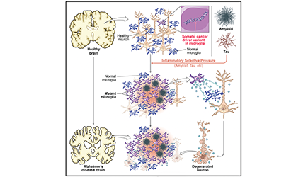
A New Somatic Genetics of Alzheimer Disease
September 3, 2026
What if every cell in your brain had its own unique genome that permanently records its history, its sibling cells, its age, the effects of disease, and its struggle for survival? How would that change the way we think about aging and degeneration in the brain? Christopher Walsh discusses these questions in light of two new studies from his lab and collaborating groups.
Community Stories
September 2, 2026
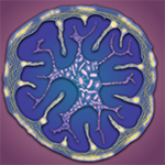
Host-microbe interactions are essential for neurological function. Valentina Lagomarsino shares new research from the Rao Lab showing how microbes play a vital role in regulating the neuroendocrine axis and activity of motor circuits in the colon.
Original article in: Nature Neuroscience >
A Shared Mechanism for Cognitive Restoration and Seizure Suppression in Neurodevelopmental Disorders
August 25, 2026
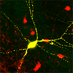
Amar Sahay shares his lab’s discovery that many ultra-high confidence risk genes for autism, bipolar disorder, schizophrenia, and epilepsy may be regulators of experience-dependent plasticity of inhibitory neurons in the hippocampus of adult mice. Proof-of-concept experiments reveal that targeting one of these regulators can improve cognition and suppress seizures in a common mouse model of neurodevelopmental disorder risk.
Original article in: Nature >

August 10, 2026
Nuria Domínguez-Iturza and Paola Arlotta present new research describing the molecular signals that promote myelination in the cerebral cortex and show that these signals are expressed at varying levels across neuronal types. Their findings reveal how neuronal diversity shapes myelin development, providing new insights into brain development and disease.
Original article in: Developmental Cell >
March 19, 2026
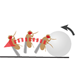
Kenichi Iwasaki shares the highlights of two recent studies exploring the use of fruit flies in research probing the neural mechanisms of object interaction. This work includes the development of a behavioral study paradigm where fruit flies are guided to interact with a small plastic ball and found to manipulate it with great dexterity.
In the News

October 9, 2026
A new strategic research initiative is seeking to bolster pioneering Harvard work in areas ripe for breakthroughs, including neuroscience, immunology, quantum, artificial intelligence, and energy and climate. The $150 million initiative, which represents one of Harvard’s largest direct investments in research, will fund promising avenues of inquiry in both basic and applied science, and fuel the creativity of the University’s exceptional scientists and researchers.
October 9, 2026
Yasuko Isoe and colleagues in the lab of Florian Engert have developed a web-based brain atlas called FishExplorer that lets researchers directly compare brain anatomy, gene expression, and neural activity across different fish species.
Original article in: Nature Communications >

October 9, 2026
Cerebral visual impairment (CVI) is a disorder caused by damage to the parts of the brain that process vision. It’s most common in babies and young children, but can continue into adulthood. In a Q+A with Optometry Times, Lotfi Merabet discusses what his research on motion processing reveals about the neural roots of CVI and where diagnostic tools like neuroimaging and eye tracking are headed.
Awards & Honors

October 9, 2026
Round up of awards and honors earned by the HBI community.
September 15, 2026
Round up of awards and honors earned by the HBI community.