by Aaron Kuan
A major quest in neuroscience is to chart all of the neurons in the brain and their synaptic connections. Such a map, called a connectome, reveals how information can flow in neuronal circuits. Obtaining connectomes is challenging because it requires imaging large volumes with high resolution. Electron microscopy (EM) is often used for high resolution imaging, but typically only records a microscopic field of view. Thanks to recent efforts to automate and speed up EM, it is now feasible to image a complete fruit fly brain, but human or even mouse brains are still way too large for comprehensive EM imaging.
In search of a faster method for circuit mapping we teamed up with scientists at The European Synchrotron in Grenoble, France, to apply a state-of-the-art imaging technique, X-ray nano-holography (XNH), to neuronal circuits. XNH uses an extremely bright and coherent X-ray beam – produced by a stadium-sized particle accelerator – to image the 3D structure of samples with high resolution.

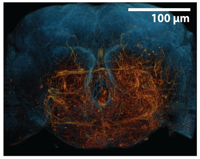
3D rendering of a fruit fly brain generated through x-ray holographic nano-tomography (XNH). The tissue outline is shown in blue, while neurons are highlighted in orange.
We demonstrated that XNH can quickly image volumes much larger than what’s accessible with EM, but still with sufficient resolution to map neuronal wiring. For example, we imaged an intact fruit fly leg and mapped how individual motor neuron axons travel between the central nervous system and the leg muscles they innervate. We also comprehensively cataloged the sensory neurons carrying mechanosensory signals from the leg.
Currently, XNH can resolve large-diameter neuronal branches, but not the finer branches or synaptic connections. However, XNH imaging is non-destructive, so we were able to combine it with post-hoc EM to resolve these fine structures as well. Using this approach in the mouse cortex, we discovered a population of neurons close to the brain’s surface (superficial layer II) in posterior parietal cortex that have unique connectivity statistics, which may underlie a special functional role.
In August 2020, the European Synchrotron completed a major upgrade, and now is the world’s first 4th generation high-energy synchrotron. In future work, we will leverage the improved quality of this X-ray source to push the limits of XNH in terms of speed and resolution. This would provide powerful new capabilities for mapping connectomes and comparing them in health and disease, from different individuals, across developmental stages, and between species.
Aaron Kuan is a postdoctoral fellow in the Neurobiology Department at Harvard Medical School, working in the lab of Prof. Wei-Chung Lee.
This story also appears in the HMS Neurobiology department newsletter, The Action Potential.
Learn more in the original research article:
Dense neuronal reconstruction through X-ray holographic nano-tomography.
Kuan AT, Phelps JS, Thomas LA, Nguyen TM, Han J, Chen CL, Azevedo AW, Tuthill JC, Funke J, Cloetens P, Pacureanu A, Lee WA.Nat Neurosci. 2020 Sep 14. doi: 10.1038/s41593-020-0704-9. Online ahead of print.
News Types: Community Stories
