We are excited to announce the winners of the 2020 Beauty of the Brain image contest! Each winner received $200. This year we received 23 submissions, of which the below five images were selected. Thank you to all who took the time to send us your images and to the 146 people who voted for their favorites. A gallery of this year’s submissions can be found here. Full image descriptions are visible if you click on the “I” information symbol on the corner of the image.
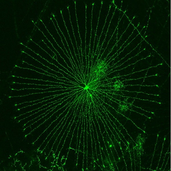
Wheel in Chick Retina
Birds have one of the most sophisticated visual system among vertebrates. Our recent cell atlas of the chick retina identifies >136 cell types. Transposon-mediated labeling with fluorescent proteins reveals the cellular diversity of the chick retina.
Masahito Yamagata
Postdoctoral Fellow
Lab of Josh Sanes, Harvard
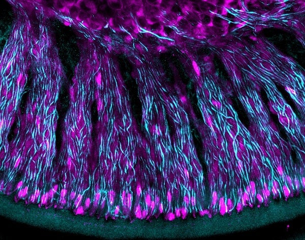
Glia and Neurons in the Inner Ear
These long, organized wires are from spiral ganglion neurons which communicate all sound information from the ear to the brain. The cells in between the wires are glia. Glia are critical for the development and organized wiring of the spiral ganglion neurons and therefore, for proper hearing function.
Isle Bastille
Graduate Student
Lab of Lisa Goodrich, Harvard Medical School
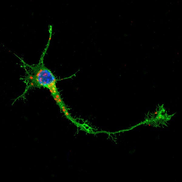
Trafficking of RNA Molecules to the Growth Cone
A young cortical projection neuron in culture with red-labeled RNA molecules being trafficked down to the growth cone, the outermost tips of the growing axons, which develop into the signaling synapses. (green: membrane-bound GFP, blue: cell nuclei, red: mRNA molecules coding for ribosomal protein, Rplp0).
Kadir Ozkan
Postdoctoral Researcher
Lab of Jeffrey Macklis, Harvard
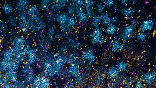
Gray Matter Galaxy
Glial cells in the temporal cortex of a human brain with Alzheimer’s disease. The star-shaped cells are protoplasmic astrocytes, which support neurons and wrap synapses with their processes, visualized here via EAAT1/EAAT2 (blue) and glutamine synthetase (gold). Meanwhile, the brain’s innate immune cells, called microglia, are stained via IBA1 (magenta).
Ayush Noori and Clara Muñoz-Castro
Undergraduate, Graduate Student
Lab of Alberto Serrano-Pozo, Mass. General Hospital
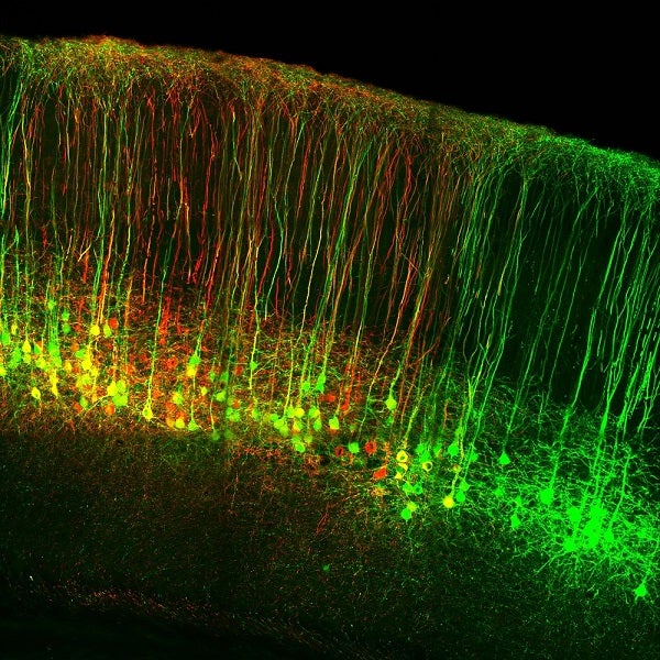
Cervical- and Lumbar-Projecting Corticospinal Neurons
Corticospinal neurons are the only direct output from the cortex to the spinal cord, providing an “information highway” to control voluntary motor function. These corticospinal neurons were labeled via retrograde viral injection into the cervical (GFP) and lumbar (RFP) spinal cord.
Carla Carol Winter
Graduate Student
Lab of Zhigang He, Boston Children’s Hospital
News Types: Awards & Honors
