
The (Bio)Physics of How Brains Fold (and Misfold)
By L. Mahadevan
One of the most striking features of the human brain is its folded surface. These folds—known as gyri and sulci—allow the expansive outermost surface layers of the brain, the cortex, to fit inside the skull. But where do these folds come from? (no, they are not forced by the skull, as known from ancient experiments too gruesome to even contemplate!) And how do genes encode developmental rules that give rise to functional geometry?
We explored this question by combining biology, physics and mathematics. We first studied how brains fold during development in three very different mammals: ferrets, macaques, and humans. Despite their differences in size, shape, and evolutionary history, all three species develop folds as their outer cortical layer expands (Choi et al. 2025).
To understand this process, we built physical models of developing brains. Starting from MRI scans of fetal or newborn brains, we created soft gel replicas with two layers, representing the cortex and the deeper brain tissue. When we placed these models in a solvent, the outer layer swelled more than the inner layer, mimicking the expansion of the cortex that is known to drive this process in vivo. The physical mimic leads to surfaces that spontaneously folded into patterns that closely resembled real brains.
These experiments suggested that brain folding might arise from a surprisingly simple mechanical principle, which we had elucidated a few years ago (Tallinen et al. 2016). As the cortex grows faster than the tissue beneath it, compressive stresses build up. Eventually the surface becomes unstable and buckles, creating folds.
To test this idea further, we developed computer simulations that model the brain as a growing soft material. Using only the geometry of the developing brain and the relative growth of the cortex, the simulations reproduced many of the folding patterns seen in real ferret, macaque, and human brains. We then used mathematical tools to compare the shapes of real brains, simulated brains, and gel models—finding strong agreement across all three approaches.
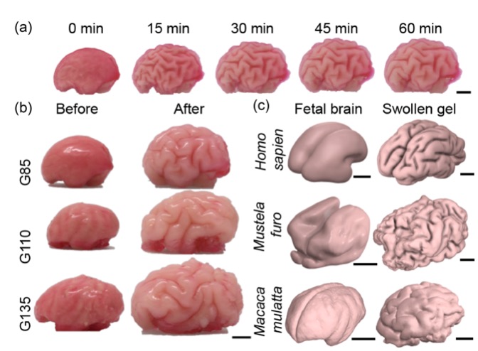
A soft physical gel model of a brain folds as it swells, and recapitulates the growth-driven morphogenesis mechanism across phylogeny and developmental stages.
(a) A gel model of a macaque brain, made from a scan at gestation day 110, wrinkles into folds over one hour (0, 15, 30, 45 and 60 minutes) as its outer layer absorbs the solvent hexane and expands.
(b) Gel models of the macaque brain at gestation days 85, 110 and 135, before and after swelling.
(c) Fetal or newborn brain scans of a human (Homo sapiens), a ferret (Mustela furo) and a macaque (Macaca mulatta), each next to the folded gel made from it. Scale bars: 1 cm
Building on this work, we then wanted to understand what happens when development goes wrong. In a companion study using ferrets, we examined genetic mutations that are known to cause cortical malformations in humans (Yin et al. 2025). Instead of trying to focus only on molecular details, we asked how these mutations change two key features of development: the thickness of the cortex and how rapidly it expands. Small changes in these quantities produced dramatically different folding patterns, including abnormalities resembling human developmental disorders such as polymicrogyria and lissencephaly. Again, our mathematical and computational frameworks and physical gel experiments that modulated these developmental features were sufficient to capture the biological phenotypes.
Together, these studies reveal how genes, cells, and physical forces interact during brain development. Genes influence how quickly cells proliferate, migrate, and grow. Those cellular processes change the geometry and growth of the cortex. Physical forces then transform those developmental changes into the folds we observe in the mature brain.
More broadly, this work illustrates how complex biological forms can emerge from simple physical principles. By combining MRI data, laboratory experiments, computer simulations, and mathematical analysis, we can begin to connect genes to tissues, tissues to organs, and ultimately form to function. Understanding how brains fold may help us better understand developmental disorders today—and perhaps even shed light on how the human brain evolved its remarkable complexity over millions of years.
L Mahadevan is de Valpine Professor of Applied Mathematics, Physics and Organismic and Evolutionary Biology at Harvard University, and Faculty Dean of Mather House at Harvard College.
Learn more in the original research articles:
Biophysical basis for brain folding and misfolding patterns in ferrets and humans.
Choi GPT, Liu C, Yin S, Séjourné G, Smith RS, Walsh CA, Mahadevan L. Elife. 2025 Dec 29;14:RP107141. doi: 10.7554/eLife.107141.
On the growth and form of cortical convolutions,
Tallinen T, Chung JY, Rosseau F, Girard N, Lefevre J, and Mahadevan L. Nature Phys 12, 588–593 (2016). https://doi.org/10.1038/nphys3632.
Morphogenesis and morphometry of brain folding patterns across species.
Yin S, Liu C, Choi GPT, Jung Y, Heuer K, Toro R, Mahadevan L. . Elife. 2025 Dec 29;14:RP107138. doi: 10.7554/eLife.107138.
News Types: Community Stories