We and other primates see more sharply than other mammals. For illustration, consider painting stripes across the moon. As many as thirty would be clear to us. This level of detail is blurred for cats, which see a uniform disk unless the stripes were so wide that the moon fit only a few. Mice are even less detail-oriented. To fit just one stripe of discernible width, the moon must be multiplied in size. What is the origin of our heightened visual acuity?
Primates see detail using a specialization of the retina lacked by other mammals, the fovea. Outside the fovea, in the peripheral retina, one is legally blind. Indeed, macular degeneration devastates vision by destroying the fovea. We sought insight into how structure and function relate in the fovea to support high-acuity vision.
A key feature of the fovea is its complement of cones, cells that initiate vision by turning the play of light into bioelectrical patterns. Foveal cones are narrow and densely packed, forming an ultrafine pixel array. By contrast, peripheral cones are broad and sparse. From this perspective, foveal cones are shaped for visual acuity. At the same time, foveal structure requires long cones. This is potentially troublesome because cones produce electrical signals at one end and transmit them at the other, 500 microns away. Indeed, foveal cones are so narrow and long that electrical signals are predicted to dissipate almost entirely in travel. How could visual performance be so exquisite with such a poor origin?

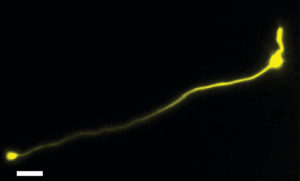
A foveal cone. The inner segment (right, attached to the soma) was injected with a dye, which filled the axon (middle) and presynaptic terminal (left). Visual information flows through foveal cones with high fidelity, due to the specialized electrical properties of these neurons and consistent with the extraordinary visual performance of primates. (Image by Gregory Bryman)”
We found that the origin is actually extraordinary. Our measurements indicate that signals traverse foveal cones briskly and with little loss. Amplification is used by textbook neurons for this purpose, but foveal cones do not need it. Instead, they have become nearly optimal conductors of bioelectricity: little current escapes them, and current within them flows with ease. Thus, the signal to be transmitted is a high-fidelity copy of the original. Cellular and behavioral performance appear aligned.
In addition to its cones, a distinguishing feature of the fovea is its miniscule size—a fraction of a millimeter. Why not have a larger fovea to see more of the world in detail? The performance of foveal cones suggests an explanation. Allow us the use of yo-yos to explain. Imagine gathering yo-yos by their strings and letting the spools rest in a single layer. The bundle of gathered strings has a smaller diameter than the array of spools. Hence, yo-yos in the middle drop straight down and have the shortest strings, while yo-yos at the margins must be displaced past the central spools and bear longer strings. Adding yo-yos means having longer yo-yos. Foveal cones, being larger at one end, follow the same rule. A larger fovea entails longer cones.
If foveal cones were extended past the lengths found in nature, our work suggests that the transmitted signal would drop in fidelity. Because we found that bioelectrical conduction is quite optimal, there is no ready way to lessen this drop. Thus, the limits of bioelectrical conduction may be one reason why the fovea is so small, and why its size is relatively constant across primates that vary from the marmoset, which stands just over seven inches high and sees with small eyes, to humans. Meanwhile, the briskness of bioelectrical conduction endows the first steps in primate vision with remarkable fidelity.
Michael Do is Associate Professor of Neurology at Harvard Medical School and Boston Children’s Hospital. The first and second authors on this study are Gregory Bryman and Andreas Liu.
This article is also featured in the HMS Neurobiology Department newsletter, The Action Potential.
To learn more, read the original research article:
Optimized Signal Flow through Photoreceptors Supports the High-Acuity Vision of Primates.
Bryman GS, Liu A, Do MTH. Neuron. 2020 Aug 18:S0896-6273(20)30579-1. doi: 10.1016/j.neuron.2020.07.035.
News Types: Community Stories
