By Brian Kalish and Eunha Kim
The brain is highly susceptible to environmental insults during intrauterine life, as it undergoes vital developmental processes, including neurogenesis, neuronal migration, and synaptogenesis. Even minor stressors during this critical period can lead to long-lasting structural and functional alterations in the brains of offspring. Activation of the maternal immune system during pregnancy is associated with a disruption in fetal neurodevelopment and predisposes to neuropsychiatric disease in offspring, but the molecular mechanisms underlying this process are poorly understood.

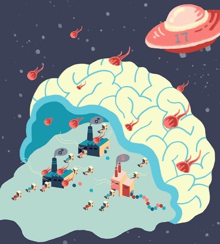
A spaceship, comets and factories denote Th17 cells, maternal cytokines and fetal brains (males vs females), respectively. Maternal inflammation disrupts translation only in male- but not in female brains.
To address this gap in knowledge, we performed single cell RNA-sequencing from the embryonic brain in a mouse model of maternal immune activation (MIA). Importantly, we profiled male and female offspring separately, given that there are known sex differences in MIA offspring. We identified cell type- and sex-specific changes in gene expression in MIA offspring. In particular, male MIA offspring demonstrated widespread changes in genes necessary for ribosome biogenesis and mRNA translation. In subsequent experiments, we found that global protein synthesis was selectively downregulated in the fetal brain of male MIA offspring.
Given this finding, we investigated signaling pathways that regulate mRNA translation, and we found that male, but not female, MIA offspring demonstrate activation of the integrated stress response (ISR). The ISR is an adaptive mechanism that can be activated under physiologic or pathologic conditions to execute a global reduction in protein synthesis as a means to shift resources towards cell survival. ISR activation was dependent on the production of the cytokine IL-17a in pregnant females. When we blocked the ISR using genetic or pharmacologic approaches, we were able to rescue male offspring from MIA-associated behavioral abnormalities.
Based on our results, we propose a model by which maternal-derived IL-17a triggers ER stress in the fetal cortex, thereby activating the ISR, arresting protein synthesis, and promoting chronic, unresolved stress at this critical stage of neurodevelopment. Moreover, our data suggest that there is a sex-specific vulnerability to ISR activation in the fetal brain. Future studies will investigate detailed mechanisms of female resiliency to MIA.
Brian Kalish is an assistant professor at The Hospital for Sick Children and University of Toronto. He recently completed a postdoc in the lab of Mike Greenberg, in the Department of Neurobiology at HMS. Eunha Kim is a postdoc in the lab of Jun Huh, in the Department of Immunology at HMS.
This story may also appear in the HMS Neurobiology newsletter, The Action Potential.
Learn more in original research article:
Maternal immune activation in mice disrupts proteostasis in the fetal brain.
Kalish BT, Kim E, Finander B, Duffy EE, Kim H, Gilman CK, Yim YS, Tong L, Kaufman RJ, Griffith EC, Choi GB, Greenberg ME, Huh JR.Nat Neurosci. 2021 Feb;24(2):204-213. doi: 10.1038/s41593-020-00762-9
News Types: Community Stories
