By Laura Holsen
Stress and negative emotional states exert powerful effects on critical aspects of human behavior, including sleep, reproduction, and feeding. However, these effects are not always uniform; substantial individual differences exist which determine whether cumulative, long-term impacts of stress and emotion ultimately influence vulnerability to disease states. We previously found that individuals with trait-level chronic stress exhibit varying levels of appetite and food-related brain reward processing in a controlled, non-stressful state. Based on this prior work, we wondered whether individuals with opposing behavioral traits related to emotional eating might react dissimilarly in terms of their physiological and brain response to a potent external stressor.
To answer this question, we recruited 28 healthy adults from the community to participate in a study protocol involving exposure to stress and measurement of blood (to assess cortisol, a hypothalamic-pituitary-adrenal axis hormone) and brain activation using functional magnetic resonance imaging (fMRI), a non-invasive method for assessing brain function. Approximately half of these subjects were categorized as emotional eaters: individuals who tend eat more when they experience strong (usually negative) emotions. The other half of the subjects were classified as non-emotional eaters. Participants completed two study visits. During one visit, they underwent a laboratory stress task designed to induce psychosocial stress (stress with a psychological and social component) and a robust cortisol response. During the other visit, they experienced a non-stressful control version of this task. At both visits, they had blood and anxiety ratings measured before and after the stress/control task, then completed an fMRI task during which they responded to reward or neutral cues: reward cues indicated they had a chance to win a snack point during that trial, while neutral cues indicated they would not have a chance to win a snack point during that trial (snack points could be used to “purchase” actual food after the scanning section). Based on the speed of their response to those cues, they obtained feedback indicating success or failure of food reward receipt. We then compared the emotional vs. non-emotional eaters on their cortisol levels and brain activation, particularly in three regions involved in reward processing: the nucleus accumbens (NAcc), the caudate, and the putamen.

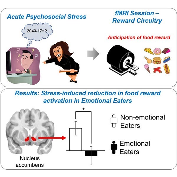
Schematic of study protocol (top) involving the acute psychosocial stressor and fMRI food reward task, and overview of results (bottom) showing reduced nucleus accumbens activation during anticipation of food reward in Emotional Eaters.
Our analyses showed three primary results. First, we saw differences in ratings of emotion and in cortisol. Emotional eaters exhibited significantly elevated levels of anxiety and increased cortisol in response to the stress task, but not the control task, while anxiety and cortisol in non-emotional eaters did not differ between tasks. Second, we observed differences in brain activation. Relative to non-emotional eaters, emotional eaters displayed reduced brain activation in the NAcc, caudate, and putamen while responding to food reward (vs. neutral) cues during the stress visit, while groups did not differ in brain activation during the control visit. Third, we found relationships between mood and brain reward activation, such that those who showed the greatest increases in anxiety exhibited the lowest levels of brain activation in the NAcc and caudate. We did not find any group differences in brain activation during the receipt component of the food reward task.
Before this study, there were no data using this approach combining a rigorous stress task vs. a control task, measurement of cortisol, and an fMRI task of food reward allowing for parsing of anticipation and receipt in emotional and non-emotional eaters. With our data, we have new insight into the mechanisms which might drive individuals to eat more during stress: a more robust HPA axis response, more anxiety, and lower activation in reward regions while anticipating food. The hypoactivation in NAcc, caudate, and putamen might trigger behaviors to compensate, such as emotional eating in an attempt to normalize reward circuitry functioning, although future studies are needed to explicitly test these trajectories temporally. Together, these aberrant responses might serve as a mechanism for maintenance of maladaptive eating behaviors during states of low mood and high stress in emotional eaters. Moreover, they point to potential wellness targets such as stress reduction to mediate unhealthy eating patterns in otherwise healthy individuals. Ongoing work in the lab will extend these findings to individuals with mental health conditions associated with disordered eating, towards discovery of novel targets for neuromodulation and pharmacologic treatment.
This work was supported the National Institute of Diabetes and Digestive and Kidney Diseases (R01DK104772; LMH, PI).
Laura Holsen is an Associate Professor of Psychiatry at Brigham and Women’s Hospital.
Learn more in the original research article:
Stress-induced alterations in HPA-axis reactivity and mesolimbic reward activation in individuals with emotional eating.
Chang RS, Cerit H, Hye T, Durham EL, Aizley H, Boukezzi S, Haimovici F, Goldstein JM, Dillon DG, Pizzagalli DA, Holsen LM. Appetite. 2022 Jan 1;168:105707.
News Types: Community Stories
