By Carla C. Winter and Zhigang He

We used viral labeling, whole-brain imaging, and single nucleus RNA sequencing to create a unified anatomic and molecular map of the neurons that connect the brain to the spinal cord.
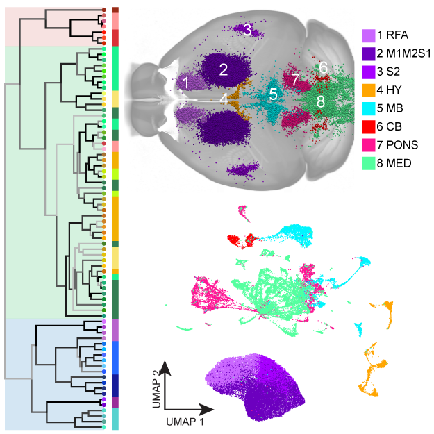
How does the brain control the body? The answer to this question, at least in part, likely resides within the neurons that connect the brain to the spinal cord. These neurons, called “spinal projecting neurons” (SPNs), act as as an information highway between the brain and the rest of the body to control movement, sensation, and autonomic functions. To increase our understanding of SPNs and how they allow the brain to carry out diverse behaviors, we, along with researchers at the Allen Institute for Brain Science, led an effort to create a comprehensive map of these neurons in the mouse brain. To do this, we used viruses to specifically label SPNs followed by whole-brain imaging and transcriptomic profiling of the labeled neurons. Whole-brain imaging allowed us to precisely map out the regions of the brain where SPNs originate from, and transcriptomic profiling revealed what genes these neurons express. In total, we profiled the transcriptomes of a total of 65,002 SPNs and computational analyses demonstrated that there are 76 different types of SPNs, each defined by a unique set of genes that they express. These computational results began to reveal their functional organization in transforming brain commands into bodily action. For example, it appears that separate types of molecularly-defined SPNs are responsible for controlling either specific parts of the body or coordinated whole-body functions, as well as modulatory gain control, which were predicted from classic motor control studies decades ago. Additionally, SPNs exhibit differential expression of genes associated with distinct firing patterns, suggesting that similar to other long projections such as visual or lower motor neuron pathways, brain-spinal cord connections might also be comprised of axons with different electrophysiological properties. By further mapping out the brain regions and gene expression profiles of these cells, neuroscientists can hope to have a better understanding of their unique functions. Looking ahead, creating this atlas will help researchers develop better therapies for pathologies that specifically damage SPNs like spinal cord injury and amyotrophic lateral sclerosis.
Carla Winter is an MD-PhD candidate in the Zhigang He Laboratory.
Zhigang He is a Professor of Neurology and Ophthalmology at Harvard Medical School and a Research Associate at the F.M. Kirby Neurobiology Center at Boston Children’s Hospital.
We are highlighting papers from Harvard labs published recently in Nature as part of of the NIH’s Brain Research Through Advancing Innovative Neurotechnologies initiative Cell Census Network (BICCN). Learn more about this initiative here.
Learn more in the original research article:
A transcriptomic taxonomy of mouse brain-wide spinal projecting neurons
Winter CC, Jacobi A, Su J, Chung L, van Velthoven CTJ, Yao Z, Lee C, Zhang Z, Yu S, Gao K, Duque Salazar G, Kegeles E, Zhang Y, Tomihiro MC, Zhang Y, Yang Z, Zhu J, Tang J, Song X, Donahue RJ, Wang Q, McMillen D, Kunst M, Wang N, Smith KA, Romero GE, Frank MM, Krol A, Kawaguchi R, Geschwind DH, Feng G, Goodrich LV, Liu Y, Tasic B, Zeng H, He Z. . Nature. 2023 Dec;624(7991):403-414. doi: 10.1038/s41586-023-06817-8. Epub 2023 Dec 13. PMID: 38092914; PMCID: PMC10719099.
News Types: Community Stories
