
How Different Neurons Shape Myelination in the Cerebral Cortex
By Nuria Domínguez-Iturza and Paola Arlotta
Complex behaviors such as dancing, playing the drums, solving mathematical problems, or speaking depend on precise communication between different regions of the brain. Many of these functions are coordinated by the cerebral cortex, the outer layer of the brain responsible for higher cognitive functions. These functions rely on billions of neurons communicating with one another through long structures called axons. Specialized brain cells called oligodendrocytes wrap selected parts of these axons in a fatty insulating layer called myelin. Much like the insulation around an electrical wire, myelin allows nerve signals to travel faster and helps precisely control their speed. This control over signal timing enables different parts of the brain to communicate efficiently and work together to perform complex tasks.
Most myelin is formed during the first years of life, a period when the brain is rapidly developing and children acquire essential motor, sensory, and cognitive abilities. However, myelination does not stop after childhood. Throughout life, experiences such as learning new skills or adapting to new environments can reshape myelin, allowing the brain to adjust communication between neurons.
When myelination is disrupted, communication between neurons becomes less efficient. Defects in myelin have been identified in a wide range of neurological and neurodevelopmental disorders, where they are thought to contribute to disease symptoms. Although myelination is essential for normal brain function, many of the signals that regulate where and when myelin forms remain unknown. Understanding these signals will help us understand why myelination goes wrong in disease and could ultimately lead to better treatments.
In this study, we investigated the molecular signals that regulate myelin development in the cerebral cortex. By combining single-cell sequencing with in-vivo gene targeting, we identified proteins that promote myelin formation when expressed by cortical neurons. Importantly, different types of cortical neurons express these proteins at different levels. These differences in expression may help shape the diverse patterns of myelination found across the cerebral cortex.
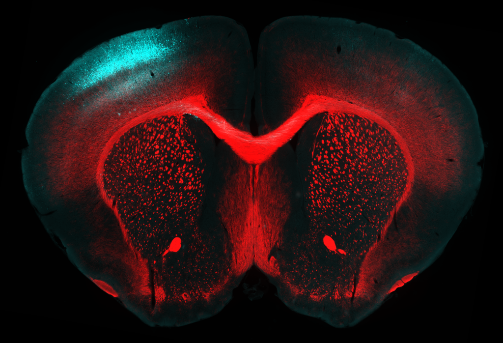
This image shows a cross-section of the brain from a young mouse. The blue cells are a group of cortical neurons that were genetically labeled before birth, allowing their visualization and the selective manipulation of gene expression to investigate the molecular signals that regulate myelin formation. The red signal marks myelin, the insulating layer that wraps axons and allows nerve signals to travel quickly and efficiently.
Why does this matter? Myelin is not distributed uniformly throughout the cerebral cortex. Instead, different types of neurons are covered by different amounts of myelin. This diversity may help fine-tune communication within brain circuits and allow them to adapt in response to experience. Understanding how these differences are established will help us better understand how the brain develops and functions, what goes wrong in neurological and neurodevelopmental disorders, and how these processes might eventually be targeted for treatment.
Ultimately, our findings suggest that different types of neurons in the cerebral cortex use distinct molecular signals to regulate how they are myelinated. This work lays the foundation for future studies investigating how the diversity of neurons across the brain shapes the development of myelin, bringing us closer to understanding both normal brain development and how myelination is disrupted in disease.
Nuria Domínguez-Iturza is a postdoctoral researcher in the laboratory of Paola Arlotta in the department of Stem Cell and Regenerative Biology.
Paola Arlotta is The Golub Family Professor in the Department of Stem Cell and Regenerative Biology.
Learn more in the original research article:
Molecular cues from distinct neuron classes drive differential myelination in the neocortex
Domínguez-Iturza N, Jokhi V, Kim K, Shetty AS, Di Bella DJ, Pereira Luppi M, Yuan W, Abbate C, Oyler-Castrillo P, Oliver NA, Venkat V, Jin X, Simmons S, Levin JZ, Brown JR, Arlotta P. Dev Cell. 2026 Jun 10;61(6):1303-1318.e8. doi: 10.1016/j.devcel.2026.05.002. Epub 2026 Jun 2.
News Types: Community Stories