By Meher Juttukonda and David Salat
The human brain is comprised of two primary tissue types: the gray matter, which is dense with neuronal cell bodies that process internal and external information, and white matter, which houses the fiber bundles that connect the different processing regions throughout the brain. Like every other organ in the body, the brain needs oxygen to generate the energy that allows it to function. Oxygen and other necessary nutrients are delivered to the brain by the systemic vascular system, which contains a complex network of blood vessels. However, the brain is unique in two important ways. First, it has a very high metabolic demand and consumes a disproportionate amount of energy relative to its size. Second, it has very little capacity to store nutrients, rendering it powerless to handle any interruptions in the supply of these nutrients. These characteristics make it imperative that blood flow to the brain remain uninterrupted.
A sudden interruption of blood flow to the brain due to a clogged artery can cause a stroke, which results in tissue death and requires urgent medical attention. While such an acute interruption in the blood supply is very serious with immediate consequences, it is becoming increasingly clear that much smaller decreases in blood flow to the brain over prolonged periods may also have serious consequences. Such chronic changes are common with aging and don’t typically present with symptoms as they occur. Instead, they may contribute to the slow deterioration of brain tissue that accumulate over the course of one’s lifetime and contribute to changes in cognitive function later in life. We used magnetic resonance imaging (MRI) to study how the delivery of blood to the brain changes with increasing age. More specifically, we used a technique known as arterial spin labeling to isolate MRI signal from the water in blood vessels to measured cerebral blood flow, or the rate of blood delivery to brain tissue.

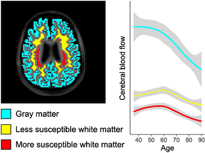
Blood flow to the gray matter of the brain (blue) is higher than in white matter but reaches its peak at younger ages before decreasing. White matter that is deeper in the brain (red) may be more susceptible to damage with aging because it receives lower blood flow than other white matter regions (yellow).
These measurements were made in a large cohort of adults at the Martinos Center for Biomedical Imaging as part of the multi-center Human Connectome Aging Project. Individuals ranging in age from 36 to 100+ years without any serious medical conditions received MRI scans of their brains and underwent other testing to determine their cognitive and overall health. The results provided us with two key findings. First, cerebral blood flow decreases with increasing age overall, but the timing of the blood flow decreases was different between gray and white matter, suggesting that there could be different processes at play in different tissues in the brain. Second, periventricular white matter regions that exhibit the lowest cerebral blood flow are also the farthest along the vascular tree. This combination of low blood flow and longer blood arrival time is intriguing as these white matter regions are known to be particularly susceptible to ischemic lesions. These lesions are common in older adults and highly prevalent in people with dementia. Our findings could be important for understanding potential vascular underpinnings of these lesions as well as their increased prevalence with aging and dementia.
Meher Juttukonda is an Instructor of Radiology at HMS and Massachusetts General Hospital (MGH). David Salat is an Associate Professor of Radiology at HMS and the Director of the Brain Aging and Dementia Laboratory at MGH. Both are faculty members in the Martinos Center for Biomedical Imaging.
Learn more in the original research article:
Characterizing cerebral hemodynamics across the adult lifespan with arterial spin labeling MRI data from the Human Connectome Project-Aging
Juttukonda, M. R., Li, B., Almaktoum, R., Stephens, K. A., Yochim, K. M., Yacoub, E., Buckner, R. L., & Salat, D. H. (2021). NeuroImage, 230, 117807. https://doi.org/10.1016/j.neuroimage.2021.117807
News Types: Community Stories
