
A New Somatic Genetics of Alzheimer Disease
By Christopher A. Walsh
Although it was long assumed that every cell in the body has the same genome, it turns out that neurons are more like fingerprints or snowflakes, with no two neurons ever sharing the exact same genome. From the moment of fertilization, the genome of the egg mutates with every cell division, so that by the time cerebral cortical neurons are formed, at 10 to 24 weeks of gestation, they inherit genomes with dozens to hundreds of mutations relative to the egg’s genome. Since these developmental mutations occur in stem cells, each mutation is inherited in a clone of cells derived from that stem cell, so that collectively these developmental mutations form a map of the unique development and clonal relationships of the brain of each of us.
But even more surprising, mutation does not stop when neurons stop dividing. Instead, neuronal genomes accumulate 15-20 new mutations each year, a mutation every 2-3 weeks, like clockwork, so that by old age normal neurons have more than a thousand mutations compared to that original genome. Two recent papers from our lab, each carried out with many amazing collaborators, show how this concept of “somatic mosaicism” in the human brain gives us new ways of thinking about the mechanisms, and possibly treatments, of degenerative brain disorders like Alzheimer Disease (AD).
The first paper looked at clonally shared mutations. We found that mutations in genes that drive cancer become more common in human brains with age. These mutations are even more prevalent in the brains of folks who died from AD, suggesting that these clonal mutations might play a role in the disease. The AD-associated mutations are found in very specific cancer genes—specifically those that drive blood cancer. And these cancer driver mutations are enriched not in neurons (which makes sense since neurons don’t divide or form cancers) but in the microglial cells of the brain, which do divide, albeit slowly.
Microglia share properties with macrophages in blood, and it seems that the cancer mutations that can push blood stem cells towards leukemia—in genes like TET2, DNMT3A, and ASXL1—cause the mutant microglia to over-proliferate and out-compete the normal microglia, so that a large fraction of microglia in the brains of many AD patients carry these cancer mutations.
The surprise is that these cancer driver mutations, when present in microglia, don’t cause tumors. Like neurons, microglia don’t form tumors. Instead, the mutations drive the microglia to an abnormal, inflammatory state, similar to what these same mutations do when they are present in blood cells. Such microglial inflammation is increasingly seen as central to how amyloid and tau ultimately cause neuronal degeneration in AD. The neurons appear to be innocent bystanders, inadvertently stuck in the middle of increasingly mutant microglia competing with each other for survival.
We are excited because the role of these cancer driver mutations opens up new possibilities in diagnostics and treatment. Clinicians may be able to identify people at risk for AD by looking for these mutations in blood (since the brain mutations are shared in blood). Also, we can consider therapeutic approaches that would re-purpose cancer drugs, designed to treat these mutant pathways, to treat AD.
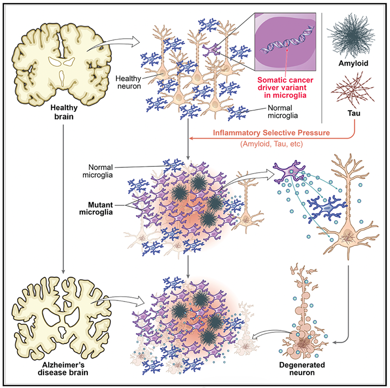
The figure illustrates schematically a working model of how age-related accumulation of cancer driver mutations occurs with normal aging (purple microglial cells), but can be accelerated in its effects by the inflammatory effects of amyloid and tau proteins in AD, causing mutant microglia to out-compete the normal microglia (blue) for survival. Mutant microglia activate inflammatory and secretory pathways that have been shown to be toxic to non-mutant microglia, as well as to neurons, potentially contributing to neuronal degeneration. Figure taken from Huang et al, (2026).
But how might mutant, inflammatory microglia actually kill neurons? Another study focusing on genomes of neurons during normal aging and in AD (and other neurodegenerative diseases as well) found that the slow, clockwork mutation accumulation seen in normal neurons with age is accelerated. One very damaging type of age-related mutation, called indels (short for insertion/deletion mutation, where a few DNA bases are removed or duplicated) accumulates very slowly normally (2-3 per year) but can be present in the thousands in neurons in AD—equivalent to hundreds of years of normal aging! The excessive, disease-related mutations suggest the presence of oxidative damage, a hallmark of inflammation, and faulty DNA repair. The increased indels appear to reflect abnormal activation of the enzyme Topoisomerase 1, which normally nicks one strand of DNA as a first step to repair it. For some reason in AD these nicks are not repaired, can reach large numbers, and are associated with fragmentation of the genome. So, it looks like the widespread genomic damage, genome fragmentation, and perhaps the death of neurons, is controlled by a process that is normally tightly regulated in neurons, but which gets badly out of control. Again, this suggests ways that we might potentially improve the condition by modulating this previously unsuspected pathway.
These two studies show how new sequencing technologies reveal our human brain cells to represent billions of individuals, each with its own genome, its own lineage, its own history, and its own individual fight for survival, competing with its neighbors. And it shows how approaching human disease by exploring this underlying genomic diversity gives us whole new ways of thinking about familiar diseases.
Christopher A. Walsh is the Bullard Professor of Pediatrics and Neurology at Harvard Medical School, Director of the Allen Discovery Center for Human Brain Evolution, Chief of the Division of Genetics and Genomics at Boston Children’s Hospital and an HHMI Investigator.
Learn more in the original research articles:
Somatic cancer variants enriched in Alzheimer’s disease microglia-like cells drive inflammatory and proliferative states.
Huang* AY, Zhou* Z, Talukdar* M, Enyenihi* L, Miller MB, Chhouk B, Rosen I, Zheng M, Zhou M, Yang A, Stronge E, Durens M, Nguyen M, Choi J, Zhao B, Khoshkhoo S, Kim J, Andersen R, An Z, Cheng Y, Ganz J, Mekerishvili L, Travaglini KJ, Gabitto MI, Hodge RD, Kaplan ES, Belk JA, Landau D, Lein ES, De Jager PL, Bennett DA, Marro* SG, Papapetrou* EP, Lee* EA, Walsh* CA. Cell. 2026 Jun 11;189(12):3719-3735.e24. doi: 10.1016/j.cell.2026.03.040. Epub 2026 Apr 21. *equal contribution.
Recurrent patterns of TOP1-mediated neuronal genomic damage shared by major neurodegenerative disorders.
Zhou* Z, Luquette* LJ, Dong* G, Kim J, Ku J, Kim K, Bae M, Shao DD, Sahile B, Miller MB, Huang AY, Nathan WJ, Nussenzweig A, Park* PJ, Lagier-Tourenne* C, Lee* EA, Walsh* CA. Cell. 2026 Jul 1:S0092-8674(26)00700-2. doi: 10.1016/j.cell.2026.06.013. Epub ahead of print. *equal contribution.
News Types: Community Stories