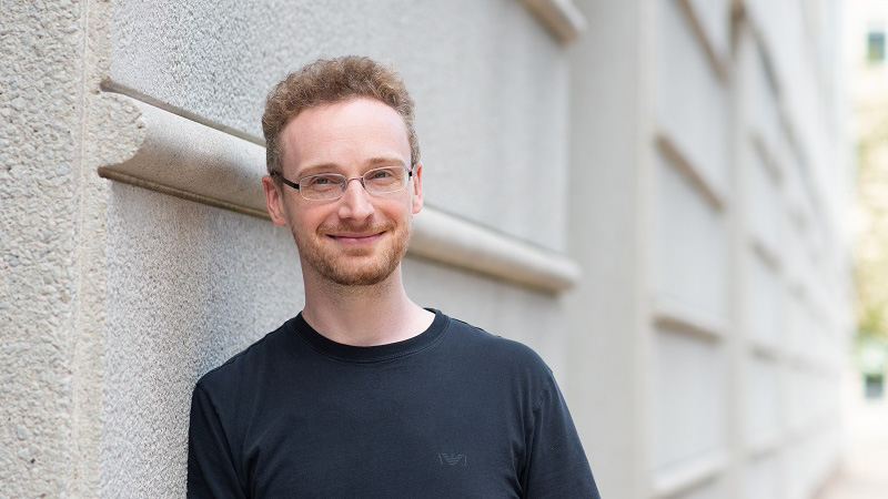
I help people solve their imaging issues and find ways to move their science forward. This usually ends up being about microscopy—how to choose which microscopes to use and the principles underlying them—but it’s also talking about the techniques to prepare a sample before getting on the microscope. In addition, it’s bringing new techniques to HMS and offering them to the research community throughout Boston.
How did you get into the neuro research world?
As a kid, I didn’t have a specific dream like wanting to be a fireman. But as I grow older, in my teens, every teacher I had felt I was too curious in a way, pestering them with lots of questions. They would say, “You should think about doing research.” And that stuck, I guess. As I finished high school, I started being interested in the brain. In France, though, where I grew up the educational system is very compartmentalized. There is no brain track. If I studied biology after high school, I would have to study every aspect of plant and animal biology broadly, and then maybe in my sixth year of studies I could do one module for a month on the brain.
So what did you do?
I tried to navigate that system for a while. One day a bunch of friends from Dallas invited me for a summer visit. Not realizing the difference in summer break between American and French colleges, I arrived there in August, just as my friends’ classes were starting. I didn’t have anything to do on my own, so I tagged along with them, sitting in on classes at the University of Texas. The first class happened to be an Intro to Psychology class that touched on the brain and neuroscience. I found out that the school had a neuroscience department with offerings for undergraduates. So I transferred there and earned my bachelor’s in neuroscience from UT Dallas.
Wow. So you so sat in on one class by chance and that changed everything?
Yes, that was really an interesting period. As an undergraduate I got interested in physiology and learned about electrophysiology and how to patch neurons. I was amazed that you could keep a brain slice alive for eight or ten hours and record activity from it.
What did you do for your graduate work?
I earned a Master’s in the UK and then went to France, earning my PhD in neuroscience and applied optics from University Paris Descartes. There I was a biologist in a physics lab that basically had never seen a brain before. They were excited to have a biologist in house and I was happy to get a crash course in physics and microscopy. Optogenetics had really started exploding around 2008 and 2009, when I joined the lab. The idea of my PhD project was to do optogenetics with two photon excitation. This had never been done before and after many trials, we used advanced optical techniques to achieve it. We were one of first labs to prove that you can actually do it.
So what is two photon optogenetics?
In optogenetics, you express light-sensitive ion channels for example in a population of cells and then shine a light that activates those cells—often in an effort to figure out what they do and what they are connected to. Generally, all of the cells that you shine light on that have the light-sensitive protein will respond at the same time. But that is far from what happens physiologically—so we designed a technique to target individual neurons and play with the sequence of activation or the number of cells necessary to get an effect.
Two photon excitation enabled us to do this, in addition shaping the light only to the areas where we wanted it to go. So you might have 100 neurons in your field of view, but be able to target a single neuron at a time. And to switch which neuron you target on the order of milliseconds. People have equated this to “playing the piano” with the brain, because you are activating whichever neuron you like with the touch of your finger.
So what brought you to HMS?
I first came here to do my postdoc in Bernardo Sabatini’s lab. The idea was to get back to doing biology while using the microscopy techniques I learned during my PhD. I built one of those fancy microscopes to study connectivity in the brain and later designed a 3D printed lightweight microscope that can be implanted on the head of mice to record neuronal activity.
What’s the coolest thing you’ve seen in your imaging facility?
It’s very specific. There’s a transgenic line in mice called RBP4, for retinal binding protein four, which is present in layer five of the cerebral cortex. Usually it’s tagged with some fluorescent protein like GFP. We cleared an entire brain like this and even by eye you could see layer five being greenish. And then when you looked through a light sheet microscope, it was blazing green. You could actually see through the whole brain, since it was cleared, and then see where these green cells fit in within. I think the most striking images are often found in extreme situations like this, where you are examining a system at the macro scale or at the nanoscale.
What is one of the challenges in your job?
Microscopy is one of those worlds where you have to make compromises and careful decisions about what you need to prove your hypothesis. There are limits to what you can do even if researchers are trying to break those limits as we speak. New techniques come up constantly. The team at NIF makes sure the core users know the current limitations and advises on alternatives. It can be difficult to impose limits on researchers’ dream experiments, but we want to help design the best scientific experiment for our users.
It sounds like you’ve traveled a lot. Do you enjoy that?
Yes, I love traveling. I’ve been to every continent besides Antarctica. I’ve been all around Europe, since that’s where I came from. Here I’ve been to Mexico. In Africa, I was mostly in North Africa—Morocco, Tunisia. I also went into the Indian Ocean, to a French island there. To Japan, where I have an aunt. And to Australia. I really love Australia.
What’s next?
Japan and Australia again if I can find the time. Also, I have a friend who moved to Hawaii recently so I may go there. I’ve always loved volcanos—I love to climb them and understand how they work.
Profile photo by Celia Muto

