Harvard Medical School
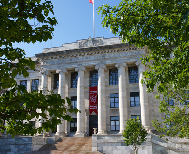
Gordon Hall, the iconic focal point of the Harvard Medical School Quadrangle, was designed in 1904 by Shepley Bulfinch (then Shepley, Rutan, Coolidge).
HBI Meetings
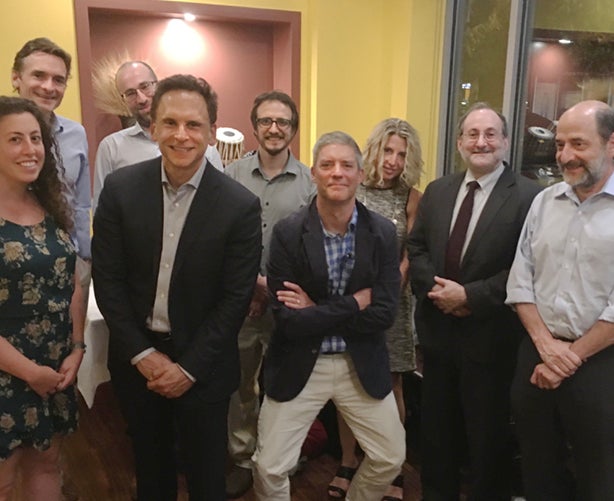
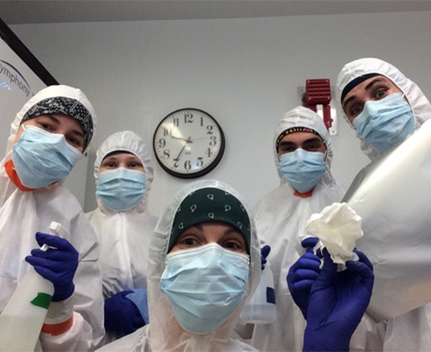
HBI developed dinner-seminar “salons” that invite investigators from diverse disciplines to share emerging findings and stimulate new ideas and collaborations. Photographed after a discussion on schizophrenia in October, 2017 it includes (clockwise, starting upper left) Steve McCarroll, Daniel Hochbaum, Beth Stevens, David Silbersweig, Mike Greenberg, Richard Born, Jordan Smoller, Rachel Ross.
HBI Grants
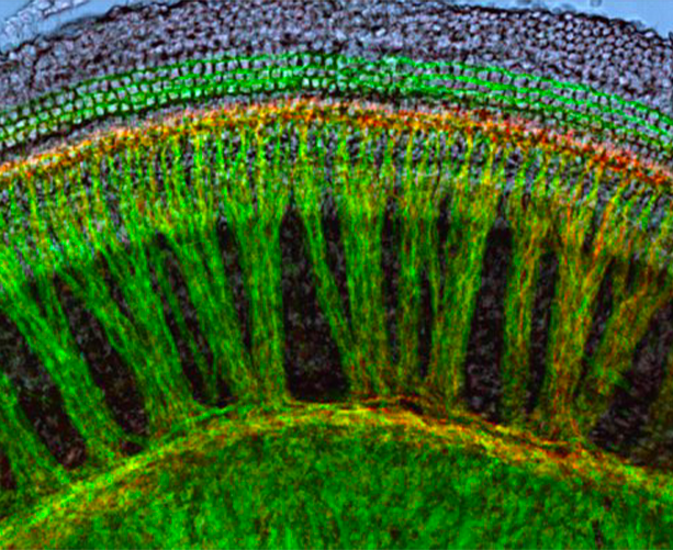
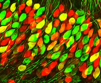
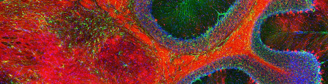
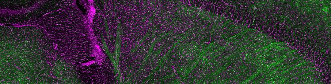
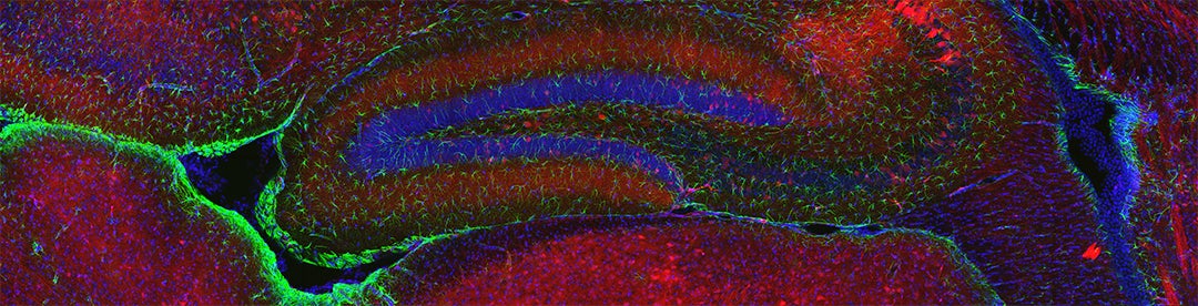
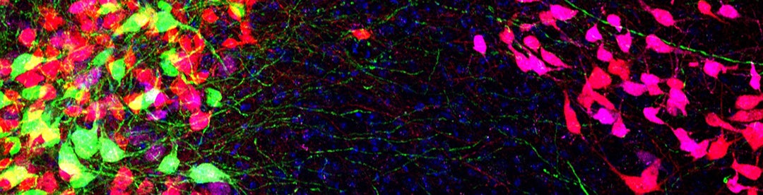
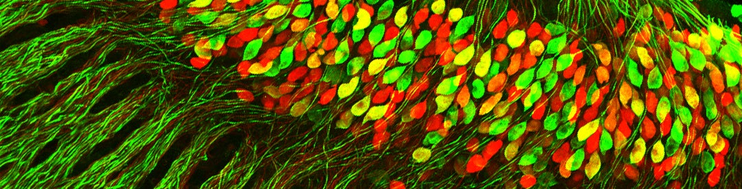
Distributed across our site are images using the Brainbow technology, a method using fluorescent proteins to enable visualization of individual brain cells in live animal models, first developed by Harvard’s Joshua Sanes and Jeff Lichtman (Livet, J. et al in Nature. 2007 Nov 1; 450 (7166):56-62.). This Brainbow image of the cochlea, from the Lab of Lisa Goodrich, HMS Department of Neurobiology, appears on the cover of the December 9, 2015 (35:49) issue of The Journal of Neuroscience.
News, Special Events
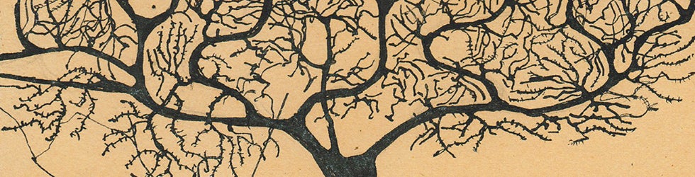
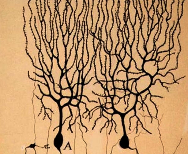
Another source of beautiful images for this site is a collection of hundreds of drawings by Santiago Ramón y Cajal (1852 – 1934), often cited as the father of modern neuroscience. The Spanish neuroscientist and pathologist shared a Nobel prize in 1906 with Italian Camillo Golgi, whose staining technique enabled and inspired Cajal’s explorations of the microscopic structure of the brain and drawings.
Our Scientists
Fun photo courtesy of the Lab of David Reich, HMS Department of Genetics
Learn Online
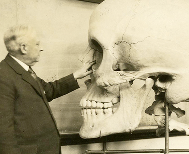
Dr. Harris P. Mosher first appointed in 1919, was Professor of Laryngology at Harvard Medical School and Chief of Laryngology at Massachusetts General Hospital. As an instructor at Harvard Medical School, he used the giant teaching skull to illustrate topics related to his specialties of ear, nose, and throat diseases. The giant skull was made in the 1890s, and is held at the Countway Library of Medicine’s Center for the History of Medicine. This and other examples of the earliest models used to teach neuroanatomy are held at Harvard by the Countway Library of Medicine’s Center for the History of Medicine and The Collection of Historical Scientific Instruments.

Design Team
Project Manager: Dana Edelman
Designer: Michael Persons, Epitome Studio
Programmer: Peter Roden
Scientist Directory Images

Scientist Directory Photo Credits
The large, customized faculty portraits were taken by Anna Olivella.