HBI Postdoc Pioneers Grant Program
Postdoctoral fellows are the lifeblood of biomedical research labs. They often develop or adapt new technologies and experimental paradigms, which allow their labs to address research questions that were previously intractable. They also can be the driving force behind a lab’s entry into a new research area. Recognizing this, HBI aims to support postdocs who take on new or risky projects.
Funded by the Office of the Provost, the HBI Postdoctoral Pioneers Program offers $50,000 fellowships to mid- to late-stage postdocs in FAS and HMS Quad Basic Science Dept labs who are conducting basic research in areas outside of their lab’s established focus. To date it has supported over 20 postdocs. Scroll below to learn more about funded projects.
We will announce a new cycle in fall 2024. Please join our mailing list to ensure you receive the latest updates.
2024 Awardees
 Aritra Bhattacherjee
Lab of Yi Zhang,
Department of Genetics, Harvard Medical School
Department of Medicine, Boston Children’s Hospital
Brain Mechanisms Potentiating Opioid Addiction Under Chronic Pain
Despite their strong addiction liability, opioids remain a principal medication for managing chronic pain. Emerging evidence indicates that chronic pain can worsen opioid addiction and its prevalence in the US is rapidly increasing. Yet, the underlying neuroadaptive mechanisms remain unclear. The prefrontal cortex (PFC) is a principal region of the reward circuit that regulates drug addiction, and also regulates pain via direct projection to the midbrain’s periaqueductal gray region. Hence PFC is believed to play an instructive role in pain/addiction interaction. However, cellular heterogeneity and functional complexity of the PFC remained a principal barrier in understanding specific molecular and circuit mechanisms that may underlie such interaction. Using single cell genomics techniques, I recently decoded the molecular, cellular and circuit organization of PFC, and identified a distinct spatially and molecularly-defined neuron subtype that responds strongly in both chronic pain and opioid taking. Anatomical tracing revealed that these neurons connect with brain regions regulating both pain as well as addiction, and these functions can be aggravated by activating the neurons. I have created a genetic mouse model to study how chronic pain facilitates addiction liability through these neurons and reveal molecular mechanisms that can promote better understanding for potential therapeutic interventions.
 Laurel Chandler
Lab of Connie Cepko,
Department of Genetics, Harvard Medical School
Visualization of Metabolism in Healthy and Diseased Retinas
The retina is a very metabolically active tissue, consuming high levels of glucose to fuel and continuously renew its light-sensing structures. It also readily shows the impact of genetic mutations, with over 300 genes leading to inherited retinal disease in humans. A common feature connecting these heterogeneous disease etiologies is that mutations that affect one cell type lead to metabolic shortcomings for others. This is exemplified in the disease retinitis pigmentosa, where mutations in rod photoreceptors lead to the secondary degeneration of cone photoreceptors. Despite not expressing the mutant gene, the degenerating cones exhibit signs of glucose deprivation and fuel starvation. However, a lack of appropriate techniques to assess the localization and levels of metabolites within the retina means the mechanism remains unclear. To address this, I aim to use fluorescence-lifetime imaging microscopy (FLIM)-based biosensors to directly measure how metabolites are consumed and shuttled within healthy and diseased eyes. I will also investigate the mechanism of several gene therapies we designed to provide metabolic support for cones and prolong vision in mouse models of retinal degeneration, which will ultimately help inform the design of future therapeutics.
 Larissa Ferrari
Lab of Isaac Chiu,
Department of Immunology, Harvard Medical School
Neurological Mechanisms Underlying Borrelia Burgdorferi-Induced Chronic Pain
Lyme disease (LD) is a tick-borne illness that is increasingly prevalent in the United States and caused by the bacterial pathogen Borrelia Burgdorferi. Pain is among the earliest neurological symptoms of LD and can persist during the illness. A major impediment to fully treat LD is an existing knowledge gap regarding the mechanisms by which Borrelia damages the nervous system. In this project, we have developed mouse models of B. burgdorferi infection where we observe long-lasting pain behaviors including mechanical and cold allodynia. We also observe induction of markers of neuronal injury and invasion of the bacteria into nervous tissues. Our goal is to understand whether the bacteria directly interact with sensory neurons to drive neuropathy and pain and identify specific brain areas activated following infection and how they intersect with regions associated with pain perception. Using a combination of transcriptional analysis, antibiotic treatment, and targeting pathways involved in nociception, we aim to identify mechanisms and changes in neural circuits that drive chronic pain in LD. Ultimately, our findings may lead to better treatments to ameliorate neurological outcomes in patients suffering from LD and its long-term effects.
 Pojeong Park
Lab of Adam Cohen,
Departments of Chemistry and Chemical Biology and Physics, Harvard University
Mapping Dendritic Voltage to Understand Single-Neuron Information Processing
Neurons transduce synaptic inputs on dendrites into axonal action potentials. Conversely, back-propagating action potentials (bAPs) traverse from soma into dendrites and modulate synaptic strength. A critical goal in neuroscience is to map the voltage dynamics throughout neuronal dendrites in live animals. Until recently, these measurements seemed out of reach, but they are now technically feasible by combining novel molecular, optical, and computational tools. I will use all-optical electrophysiology with a virtual navigation system in mice. The whole dendritic trees will be visualized using a microprism inserted into the hippocampus, a brain area critical for spatial learning and memory. This approach will elucidate the propagation of excitation, inhibition, and bAPs within dendrites, their interactions in space and time, and the influence of neuromodulatory inputs on dendritic processing and plasticity during place cell development. This study will provide new insights into single-neuron computation.
 Rebecka Sepela
Lab of Nicholas Bellono,
Department of Molecular and Cellular Biology, Harvard University
Lab of Jon Clardy,
Department of Biological Chemistry and Molecular Pharmacology, Harvard Medical School
Environmental Microbiomes Drive Chemosensory System Evolution
Three billion years after bacterial life originated, animals diverged from their protistan ancestors and began to evolve in a bacterial world. Captivating examples of interkingdom symbioses demonstrate genetic and physiological ramifications of animal and microbial relationships. The fundamentals of many host and internal microbe interactions are beginning to be appreciated, yet whether animals exploit omnipresent microbial cues to define their external environment is less clear. Here, I will interrogate the chemical biology of surface-specific microbial communities and then use key microbial molecules as tools to understand how animals evolve to interact with their environmental niche. One animal whose sensory system operates in close apposition to microbe-coated surfaces is the octopus, which relies on its eight arms to explore the seafloor. Using the octopus chemotactile sensory system, I will broadly ask how animals use cues from their environmental microbiomes to inform surface-specific behaviors. Decoding interkingdom communiques is a novel approach to studying (1) the environmental pressures influencing sensory system evolution, (2) the origins of symbiotic relationships, and (3) the influence of our changing environment on animal behavior. Findings from this study will inform foundational principles regarding evolution and could have ramifications in human-microbe relations for health and disease.
 Jonnathan Singh Alvarado
Lab of Mark Andermann,
Department of Neurobiology, Harvard Medical School
Department of Medicine, Beth Israel Deaconess Medical Center
Identification of Brainstem Circuits That Mediate Chronic Pain Perception
A single brain region can contain dozens of neuronal subtypes that are involved in diverse functions. Nonetheless, studies in most model organisms tend to focus on a single cell type at a time, partly due to the technical limitations of our available tools. These obstacles have prevented a comprehensive view of how multiple components of the neuronal “ecosystem” work together. To overcome these issues, we are developing a novel pipeline that will enable us to link genetic and functional information in up to thousands of neurons at a time. We are using these methods to identify pain-sensitive neurons in the brainstem that contribute to chronic forms of pain, with the goal of paving the way for new, selective analgesics that target the brain. Ultimately, we hope to share our methods with the wider HMS community, as they can be adopted to the study of any behavior or brain region.
 Aditya Venkatramani
Lab of Xiaowei Zhuang,
Departments of Chemistry and Chemical Biology and Physics, Harvard University
Multiplexed Optical Barcoding and Sequencing For Spatial Omics
The functions of tissues emerge from the combined actions of the various cells within. Spatial omics is an approach to measure the molecular profiles of tissues in a spatially resolved way. This can be used to identify cell types, map their spatial organization and interactions, interrogate their functions, and thereby provide insights into the molecular and cellular mechanisms of tissues.
Spatial omics methodologies fall broadly into two categories: sequencing-based and imaging-based approaches. While sequencing-based tools capture nucleic acids without targeting specific sequences for unbiased discoveries, their spatial resolution is often limited to larger than a single cell. Conversely, imaging-based methods offer sub-cellular spatial resolution but are a targeted method that profiles hundreds to thousands of selected genes. We are developing a method that integrates the strengths of both imaging and sequencing to offer a novel approach to spatial omics with the potential for untargeted genome-wide profiling with single-cell resolution.
Our ambition is to leverage this new method to study systems with heterogeneous cell types and gene expressions such as a developing brain and tumors. In such cases, the sample-to-sample variability is high, and phenomena such as metastasis and transient cell types during development are poorly understood. We hope to create a holistic understanding of the molecular underpinnings of such phenomena.
2023 Awardees
 Corey Allard
Lab of Nick Bellono,
Department of Molecular and Cellular Biology, Harvard University
Connecting development and physiology to understand the evolution of novel traits
How novel traits originate is a fundamental and unanswered question in biology, and research has been hampered by a paucity of suitable model systems. An ideal model to study this question would comprise a group of organisms that possess a unique, major trait that exhibits diversity among species resulting in a range of discrete functions and behaviors. One remarkable example of such a trait is the ‘leg’-like sensory appendages unique to the family of fishes known as sea robins. These unusual structures represent detached pectoral fin-rays specialized to function as sense organs that mediate sensation of chemical and tactile stimuli. Remarkably, closely-related sea robin species use their legs to facilitate dramatically distinct predation strategies, including a restricted lineage of sensory specialists which have the remarkable ability to locate and uncover prey items buried in sand. This ability is driven by sensory innovations unique to the legs of digging sea robins. Through an integrative strategy combining physiological, genetic, and behavioral analysis, we will leverage sea robins to investigate the origins of evolution novelty, and explore questions including (1) the developmental and evolutionary basis of leg acquisition and (2) the molecular and cellular basis for sensation and sensory specialization in the legs.
 Ryunosuke Amo
Lab of Naoshige Uchida,
Department of Molecular and Cellular Biology, Harvard University
Effect of opioids on dopamine control
Opioid addiction has been declared a public health emergency in the US, while medical opioids are indispensable for managing pain. Although dysfunction of dopamine is related to analgesia and addiction with opioids, the exact mechanisms remain unclear. It is widely believed that dopamine neurons convey reward prediction error (RPE), the difference between expected reward and actual reward outcome. This RPE signal is used as an effective teaching signal in machine learning. I have been studying what algorithm is used to compute RPE and how this signal is generated in dopamine neurons. In this project, I will apply the techniques and knowledge of computational and systems neuroscience of dopamine to analyze the effect of opioids on the dopamine system. I aim to reveal what information in dopamine neurons is affected by opioids and how opioids alter neural circuit function. Through these studies, I hope to uncover novel mechanisms that explain analgesia and maladaptive behaviors seen in opioid addicts, such as their inability to avoid negative consequences.
 Kiah Hardcastle
Lab of Bence Ölveczky
Department of Organasmic and Evolutionary Biology, Harvard University
Examining how basal ganglia supports the shaping of new motor behaviors
We can shape new motor behaviors through trial-and-error learning. For example, with practice we can learn to shoot a free throw in basketball. While Thorndike’s Law of Effect, which states that animals repeat rewarding movements and suppress unrewarded ones, has been a guiding principle of trial-and-error learning, this rule doesn’t fully explain learning in complex, naturalistic behaviors. For example, how do animals decide to repeat a successful behavior versus explore other variants that could be easier or more rewarding? Further, many mysteries remain when it comes to how trial-and-error learning is implemented in neural circuits. For example, with practice, motor behaviors like a free throw become stereotyped, driven by a ‘motor memory’. But how does neural activity change to support the ‘memory’ of this new behavior? In my work, I examine these questions through the lens of rodent behavior and the sensorimotor basal ganglia (BG). I leverage advances in 3D pose tracking and automated behavioral training to shape an animal’s movements into a new behavior, allowing me to measure the animal’s entire behavioral repertoire during learning. I also record from the BG during learning, providing me an unprecedented view into how neural circuits are transformed by learning.
 Carole Hyacinthe
Lab of Clifford Tabin
Department of Genetics, Harvard Medical School
Decoding the genetic basis of acoustic behavior
“Acoustic behavior”, or the intentional use of sounds for communication between members of the same species, is a common feature in the animal kingdom, including being a key element of our own daily life. Our understanding of how the brain controls acoustic behavior, and how consequent sounds are physically produced, are becoming more precise. However, the genes responsible for differences in inherited patterns of acoustic communication are practically unknown.
We recently discovered that a fish species, Astyanax mexicanus, uses sound to communicate. What makes this fish interesting is that, originating from a single species, two different subtypes of Astyanax have evolved: a sighted “surface fish” populating the noisy rivers of Mexico, and a blind “cavefish” adapted to the quiet and permanently dark caves. Both produce sounds, but their trigger, meaning and use differ. Moreover, depending on their regional location, independent fish populations display distinct “accents”. Importantly, these differences are maintained in a lab setting, indicating that they are hard-wired. The power of Astyanax is that surface and cave fish can be bred with one another and produce fertile offspring, allowing assessment of inherited acoustic behavior in offspring, and ultimately permitting the identification of genes responsible for this behavior.
 Yunjin Lee
Lab of Jun Huh
Department of Immunology, Harvard Medical School
How do gut bacteria regulate sociability?
The idea that gut microbes regulate brain function, aptly coined as the ‘gut-brain axis,’ has recently garnered significant attention. However, underlying mechanisms are uncertain due to the scarcity of suitable animal models to study this process. One needs to establish a model where gut bacteria influence sociability. We re-derived a widely used monogenic mouse model for autism, Cntnap2-KO, into a germ-free (GF) condition. Intriguingly, Cntnap2-KO mice displayed sociability deficits at a specific-pathogen-free (SPF) condition, not under a GF condition. Thus, our lab successfully established a system where bacterial colonization produces about distinct sociability phenotypes in a genetic background-dependent manner. This research will allow us to elucidate mechanisms by which gut bacteria promote autism-like symptoms by influencing brain function.
 Leland Wexler
Lab of Max Heiman
Department of Genetics, Harvard Medical School
Department of Pediatrics, Boston Children’s Hospital
A sensory cilium mediates specific neuron-glia attachment
Sensory cilia play critical roles in photoreceptors, olfactory neurons, taste cells, and hair cells. Using C. elegans, I discovered that the sensory cilium of the oxygen sensing neuron URX forms a highly stereotyped attachment to a specific glial partner, the ILso glial cell. Cilia function primarily to sense a cell’s external environment and transmission of this sensory information. A for cilia role in cell adhesion is novel direction for cilia biology. It raises the possibility that cilia function can be modulated by specific cell-cell interactions. My projects aims to investigate the possibility that the cilia-glia attachment allows the glial cell to modify the structure of the cilium and thus tunes the responsiveness of the sensory neuron. I aim to identify the molecular mechanisms establishing this cilia-glia attachment and determine the significance of this attachment for neuronal function.
 Rachel Wolfson
Lab of David Ginty
Department of Neurobiology, Harvard Medical School
Understanding the colon innervating sensory neurons that respond to physiologic and pathophysiologic stimuli
Colon sensation is critical for water resorption, motility through the gastrointestinal tract, defecation, and pain perception. The sensory neurons that innervate the colon and skin are those with cell bodies that reside in the dorsal root ganglia (DRG). While the skin innervating DRG sensory neurons have been characterized, those innervating the colon are poorly understood. Single cell sequencing has revealed a vast genetic heterogeneity within the DRG, with at least fifteen transcriptionally distinct sensory neuron subtypes, but the identity and functions of the DRG neuron genetic subtypes that innervate the colon are unclear. My goal is to define the genetic subtypes, morphologies, and response properties of colon innervating DRG sensory neurons. Characterizing the DRG sensory neuron subtypes that respond to different stimuli is necessary to understand which neuronal subtypes mediate pain responses under different pathophysiologic states, identifying populations that can be targeted for therapeutic purposes.
2022 Awardees
 Evelyn Avilés
Lab of Lisa Goodrich,
Department of Neurobiology, Harvard Medical School
DCC-mediated molecular mechanisms of axon guidance
Our abilities to move, think, and sense and respond to the environment rely on a functioning nervous system. Neurons form connections with other cells to generate circuits that transmit signals resulting in an organism’s behavior. During development, these connections form by the extension of neuronal processes, called axons, that grow toward specific target cells. Axons can travel very long distances without being derailed from their stereotypical tracks. For this guidance to happen precisely, axons respond to molecules that they encounter in the tissue. For example, the secreted protein Netrin1 can act over a long-distance to guide axons expressing the receptor DCC on their surfaces. Even though this system has been studied for many decades, how Netrin1-DCC interactions enable the variety of guidance decisions that axons and cells make during development is unknown. Draxin is another secreted molecule that can interact with DCC, possibly influencing how the same axon responds to Netrin1. We aim to investigate how the DCC receptor modulates the action of Netrin1 by collaborating with Draxin in vivo and whether discrete portions of the DCC molecule have differential roles in axon guidance.
 Ava Carter
Lab of Michael Greenberg,
Department of Neurobiology, Harvard Medical School
High-throughput study of human-specific aspects of neuronal activity-dependent transcriptional programs
The human brain displays remarkable cognitive capabilities, achieved largely through increased size and connectivity in the neocortex. Sensory experience contributes to the refinement of connectivity, in part through the action of genes that are activated in response to stimulus within neurons. We have identified an evolutionarily young family of genes, the KRAB-domain containing Zinc Finger Transcription Factors (KRAB-ZNFs) whose expression increases in human neurons following the application of a stimulus. To understand how each of these KRAB-ZNFs contributes to the cellular programs activated by neuronal stimulus, we are employing a new CRISPR-based screening technology. With this technology we can disrupt the expression of hundreds of genes in human cortical neurons, and assess the effect of their loss on gene expression in single neurons. Development of this technology in our lab will allow us to identify those KRAB-ZNFs, and other genes, whose expression is critical for driving human-specific features of brain development and response to external stimuli.
 Yasmin Escobedo Lozoya
Lab of Susan Dymecki,
Department of Genetics, Harvard Medical School
Modulating the encoding of reward memory
As a postdoctoral fellow in Susan Dymecki’s Laboratory, I explore how neuromodulatory systems can exploit the intrinsic flexibility of brain circuit dynamics to allow neuromodulatory transmitters such as serotonin to nudge circuits into activity states or behaviors useful to the animal. I study neuromodulation in the context of emotional memory formation by neurons that signal through both serotonin and glutamate.
My work proposes that a class of serotonin neurons gates the transfer of information between brain regions responsible for reward memory formation that may contribute to problematic states such as drug relapse. If correct, this model would suggest some neuromodulatory neurons can exert circuit actions akin to the switches of an old-fashioned telephone switchboard: they can control which circuits should be connected and when by tuning their dynamics to allow or prevent communication, thereby turning inter-area communication on or off.
The Pioneer Award will allow us to extend our experimental reach to record brain oscillatory dynamics and select neuron activity via in vivo electrophysiology, calcium imaging, and electroencephalography and test these hypotheses.
 Jonathan Green
Lab of Christopher Harvey,
Department of Neurobiology, Harvard Medical School
Scalable methods for understanding the cognitive roles of brain cells in the cerebral cortex
To understand how we think, we need to understand the basic building blocks of our brain – its neuronal cell types. We are developing new tools to study cell types in a more precise and scalable manner and applying these tools to study spatial cognition in cortex. Using a novel viral tool that targets a subtype of somatostatin neurons, we found that in posterior parietal cortex this subtype selectively and synchronously activates as the mouse corrects for errors as it navigates toward a reward. Using cellular resolution optogenetics, we found that these cells tend to excite each other, helping to explain their synchronous activity during error corrections. Previous studies in the same area and the same task did not identify this signal because it is present in a rare neuronal subtype, highlighting the importance of precise cell type tools. The discovery of this error correction signal is exciting because error signals are fundamental both to executing and learning tasks. My goal for the future is to build on this work to understand the functions of this error correction signal, and the functions of other cortical cell types in cognition.
 Dilansu Guneykaya Cinar
Lab of Sandeep Robert Datta,
Department of Neurobiology, Harvard Medical School
The cell type specific role of Ms4a6 genes in Alzheimer’s Disease
Alzheimer’s Disease (AD) is a complex, multifactorial neurodegenerative disorder that causes memory loss, difficulty completing tasks, withdrawal, mood changes and ultimately death. Unfortunately, current therapeutic strategies are significantly limited. Recently, genetics research drew many road maps for AD researchers revealing potential risk factors to understand the disease progression. Among the genes implicated in these studies, certain members of the Ms4a gene family are suggested to be associated with immune regulation of AD. In addition, we know that Ms4a genes are expressed widely by many immune cell populations, including brain resident immune cells, myeloid cells and lymphocytes. My current lab (Datta lab) previously identified expression of Ms4a gene family in olfactory sensory neurons and developed related mouse models. Recent studies suggested the strong connection between central nervous system (CNS) and immune system. My research project aimed to investigate the consequences of Ms4a gene manipulations in immune cells in a subset of AD mouse models. This will allow us to identify cross reactions between CNS and periphery, as well as their ability to influence the onset or progression of AD. We are planning to learn more about how the Ms4a gene family acts in different immune cells, and influence the risk of developing AD.
 Ryan Maloney
Lab of Benjamin de Bivort,
Department of Organismal and Evolutionary Biology, Harvard University
Using expansion microscopy to understand the basis of individuality
What makes an individual an individual? While genetics and environment play a significant role, even genetically identical flies raised in the same environment show a wide range of individual preferences on a number of tasks. This poses a key question: what in the nervous system (and therefore what in our behavior) is determined by genetics and observable changes in our environment, and what parts vary by chance?
Answering this question requires looking at the parts of the fly brain involved in these behaviors with extreme detail, across a large number of flies with different measured preferences. Few techniques combine the resolution and throughput required to do this, however we’ve embraced a novel technique called Expansion Microscopy that does. Expansion Microscopy allows us to embed the fly brain in a gel and literally enlarge it to get a closer look at the neurons underlying behavioral circuits. This technique allows us to look at different proteins across different neurons and see how they vary between individuals—and in doing so understand how individuality at the molecular level connects to individuality in behavior.
 Souvik Mandal
Lab of Venkatesh N. Murthy,
Department of Molecular and Cellular Biology, Harvard University
How do simple minds make complex decisions? Deciphering the “basic rules” of individual and collective decision-making in ants
I am broadly interested in how behaviors in social animals are influenced by their evolutionary history, lifetime experience (i.e., learning and memory), and recent experiences. Social insects are of special interest because with a simple brain in their repertoire, they accomplish complex tasks, involving both individual as well as collective decision-making; they do so using simple and elegant rules of thumb. My research focuses on finding these minimal rules by quantifying animal behavior using cutting-edge technologies like computer vision and artificial intelligence. Currently, I am working on understanding how ants integrate current and prior olfactory information for making decisions individually (while searching for food in their natural habitat and following pheromone trails), as well as collectively (while working together to achieve a certain common goal). While I am developing theoretical frameworks of decision-making from these behavioral strategies, these can further be translated into algorithms that could inspire the development of new solutions for computational problems in the field of artificial intelligence.
 Jeongho Park
Lab of Talia Konkle,
Department of Psychology, Harvard University
Full-field fMRI: a novel approach to study immersive vision
Traditional functional MRI setups are limited to presenting images in the central 10-15 degrees of visual field, whereas we experience a more than 180 degree view of the world every day. In this project, we have developed a new method for ultra-wide angle presentation in the scanner, enabling us to measure responses for the first time in cortex responding to the far-periphery, and to immersive first-person views of environments. This works by bouncing an image off two angled mirrors directly into the scanner bore onto a custom-built curved screen, which creates an unobstructed view of 175 degrees. Additionally, we present images with a compatible wide field-of-view, rendered from custom-built virtual environments—a critical step as using standard pictures leads to highly distorted perceptions. With this new setup, now we can stimulate parts of brain that were methodologically out of reach. Further, we can examine brain responses to visual environments in an immersive setting beyond postcard-like views of the world used in current methods. More broadly, this project will provide new empirical traction on current theories which argue that fovea-to-peripheral organization underlies the basic blueprint of the visual system and its interface to broader whole-brain architecture.
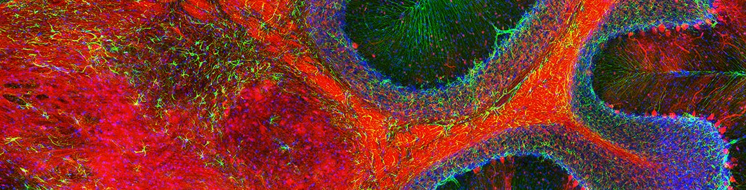
Banner image shows staining of cerebellum, courtesy of David Brann, graduate student in the lab of Bob Datta.
Most photos by Celia Muto. Photo of Yasmin Escobedo Lozoya by Rev. Mark Santucci.