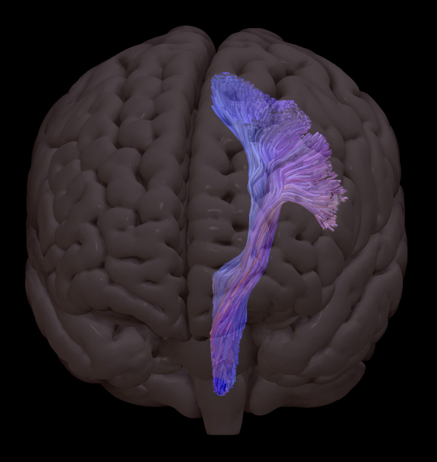
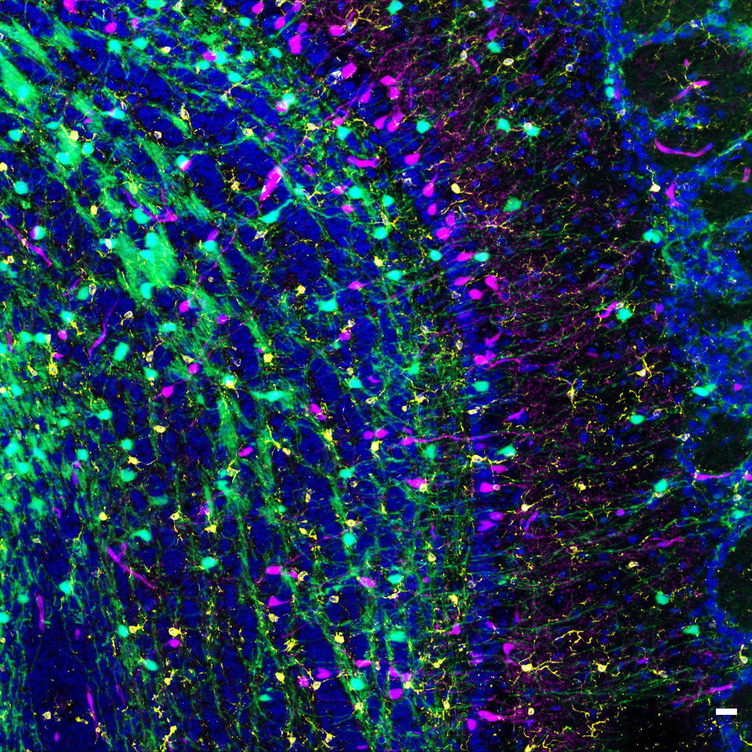
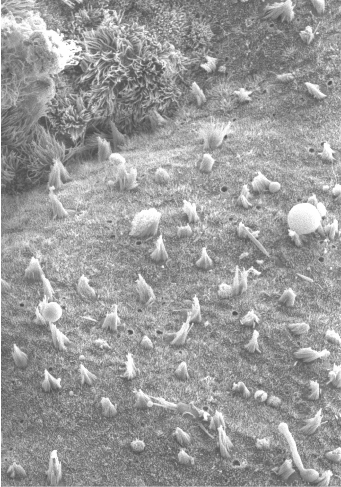
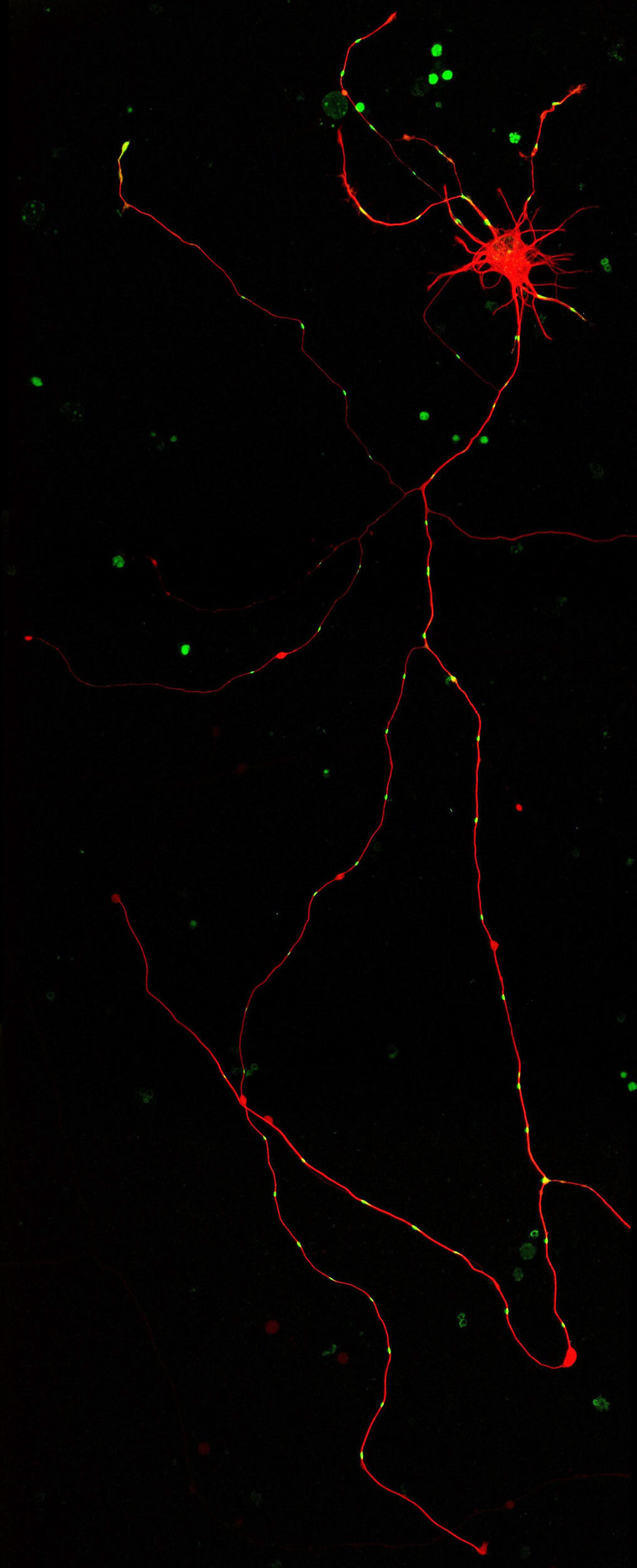
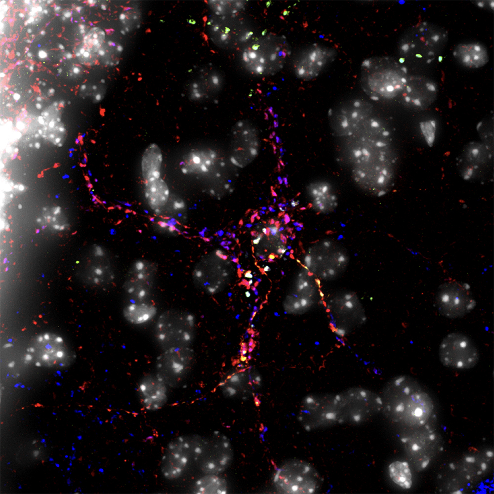
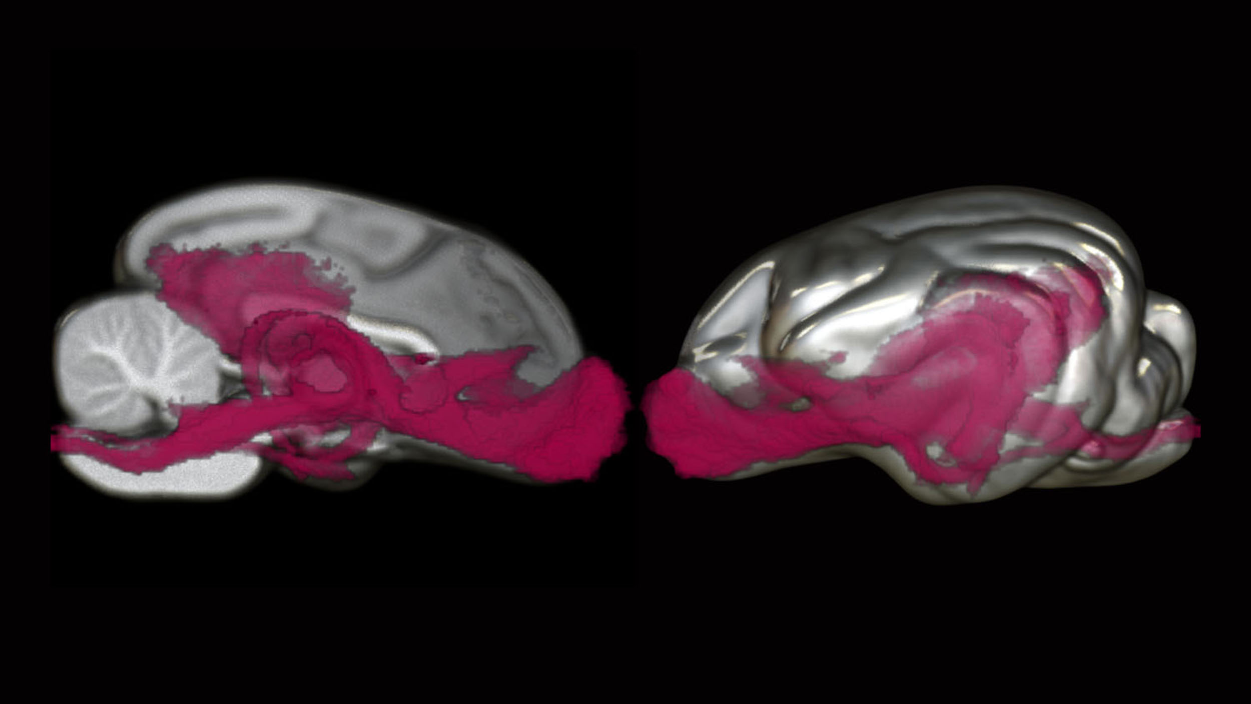
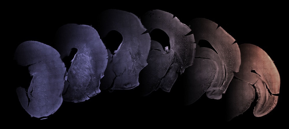
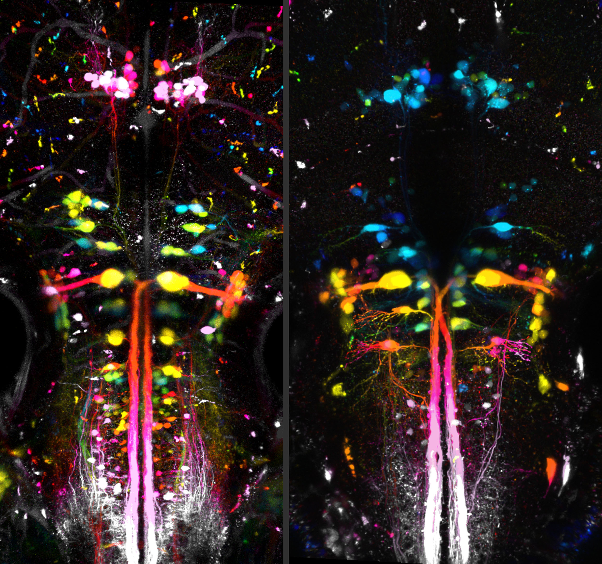
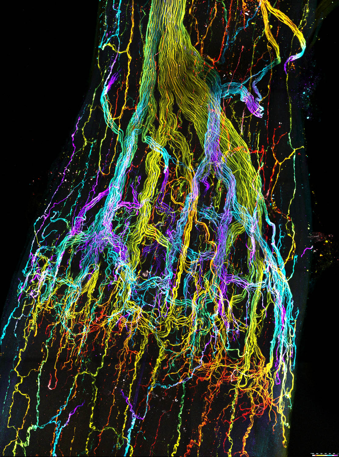
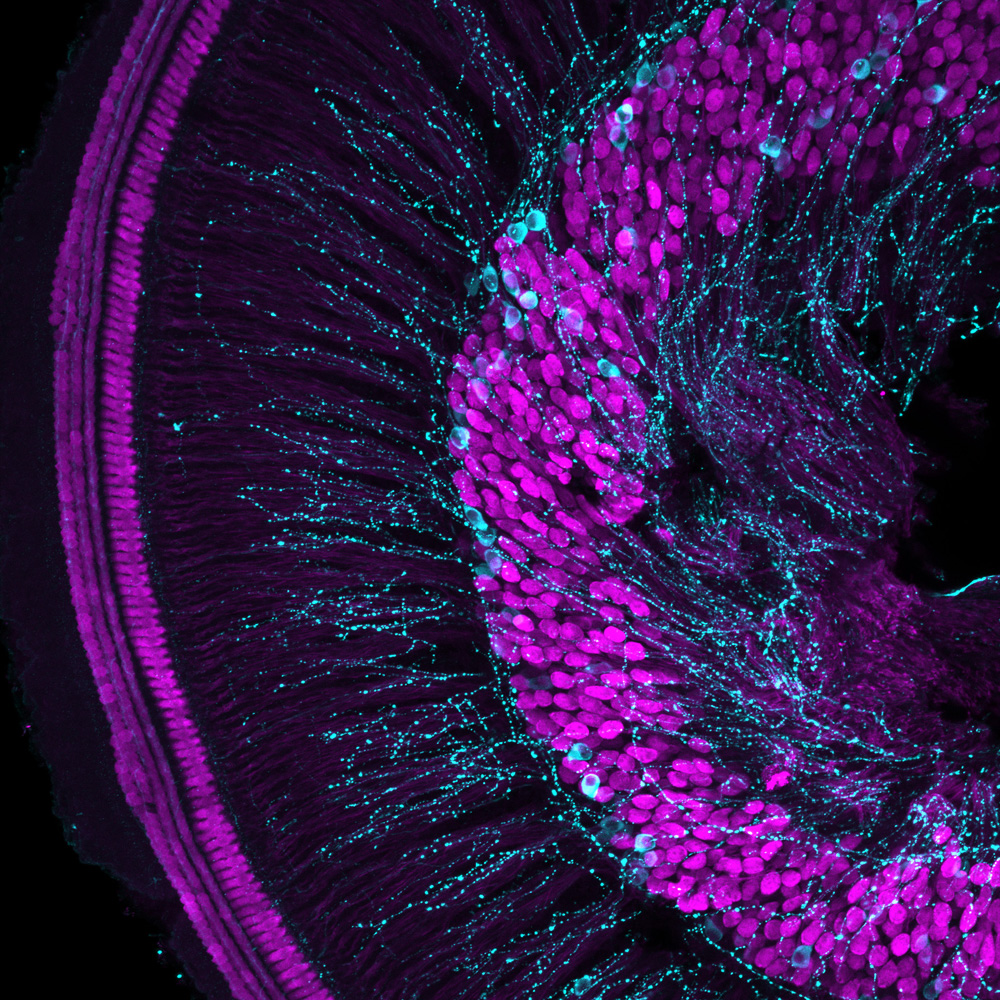
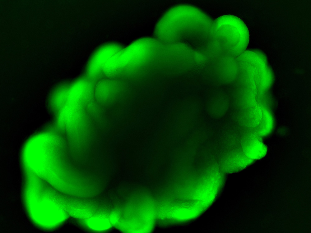
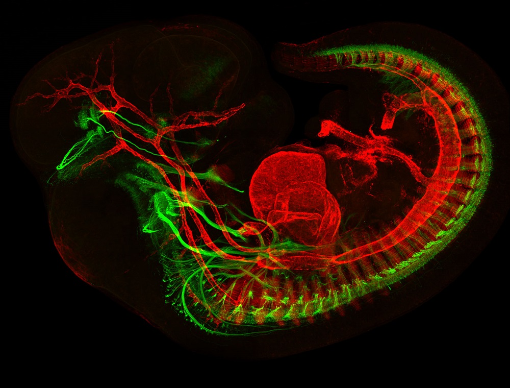
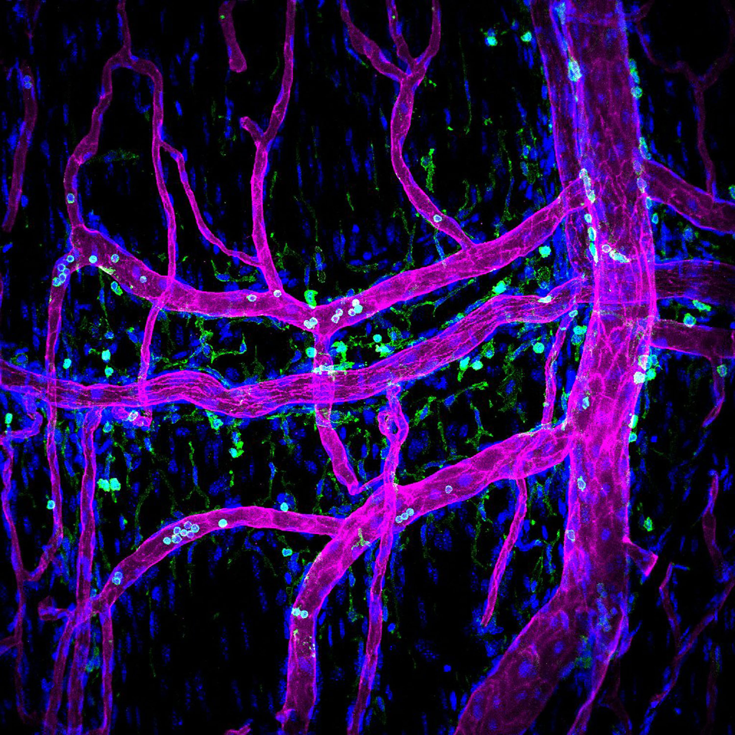
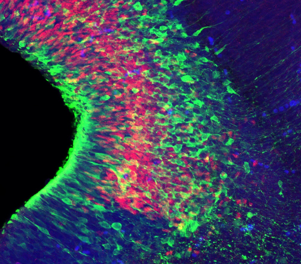
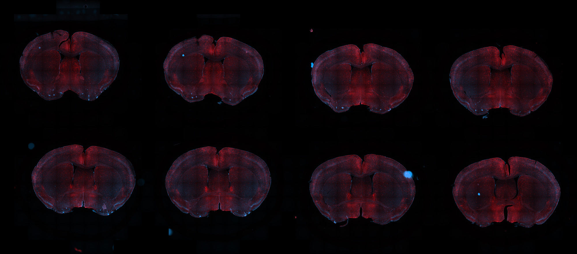
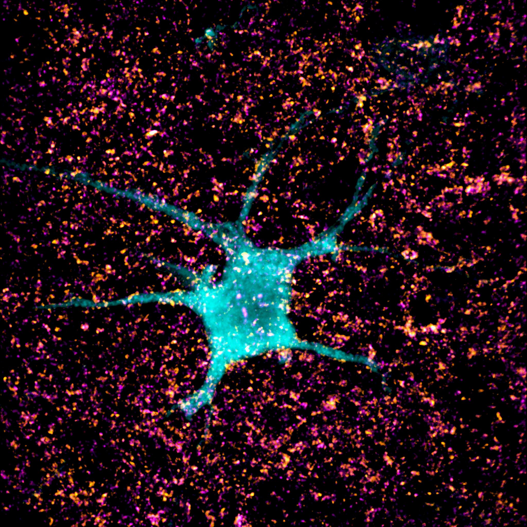
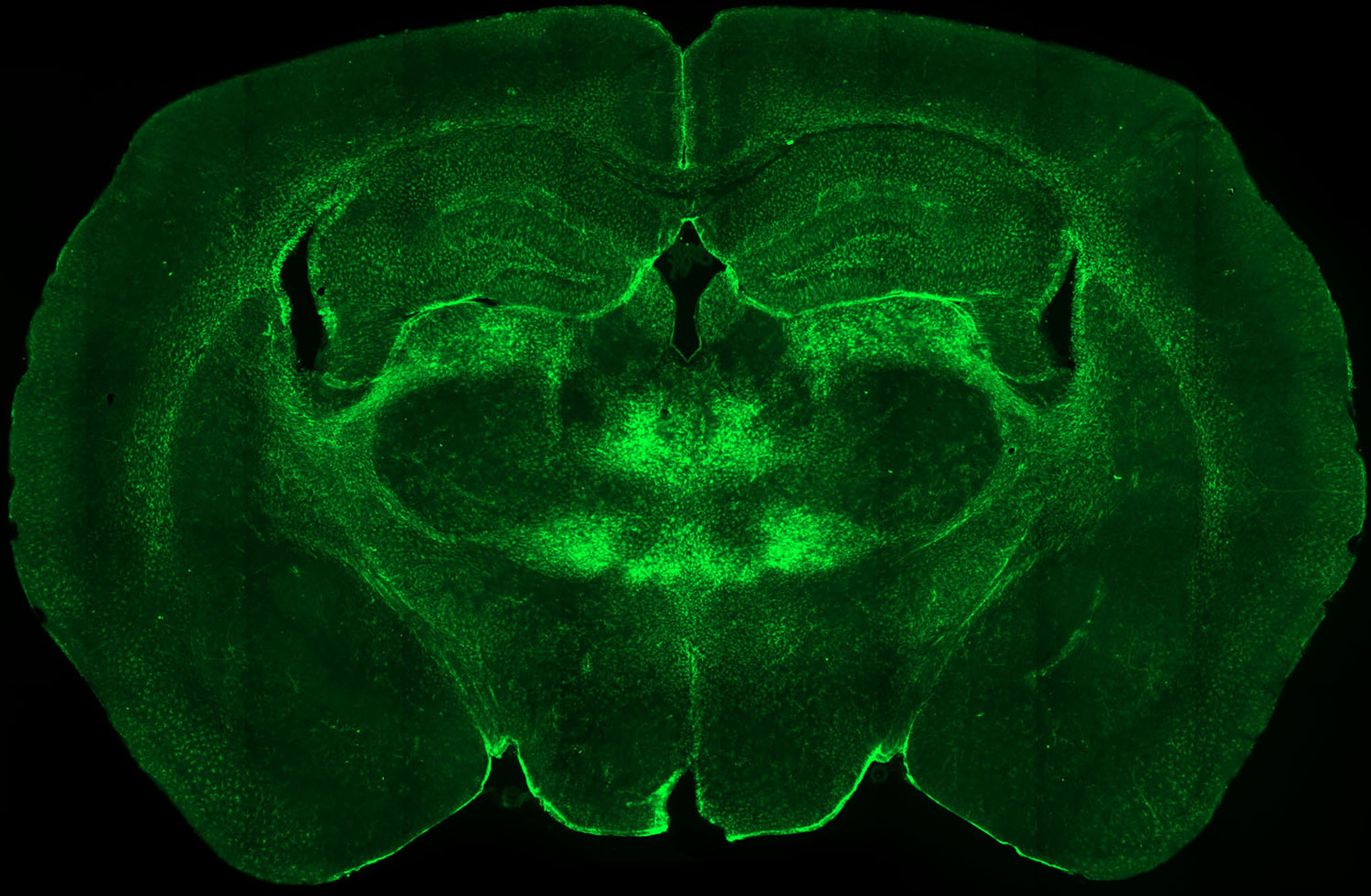
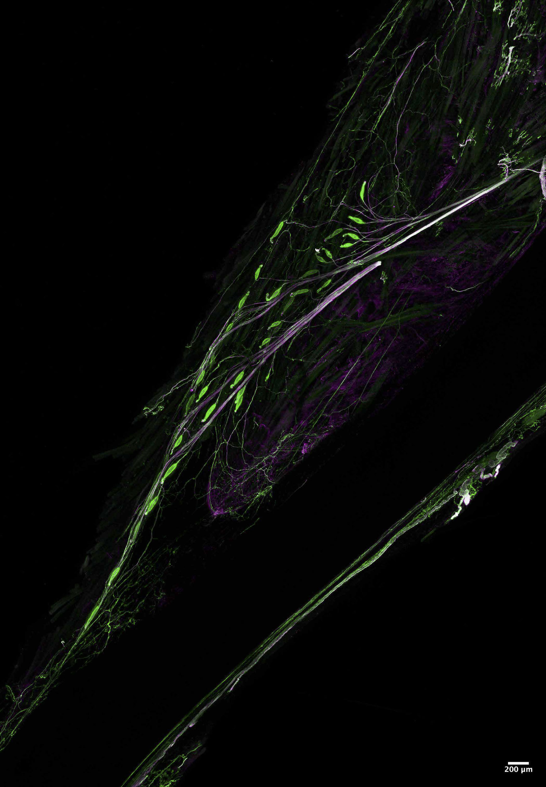
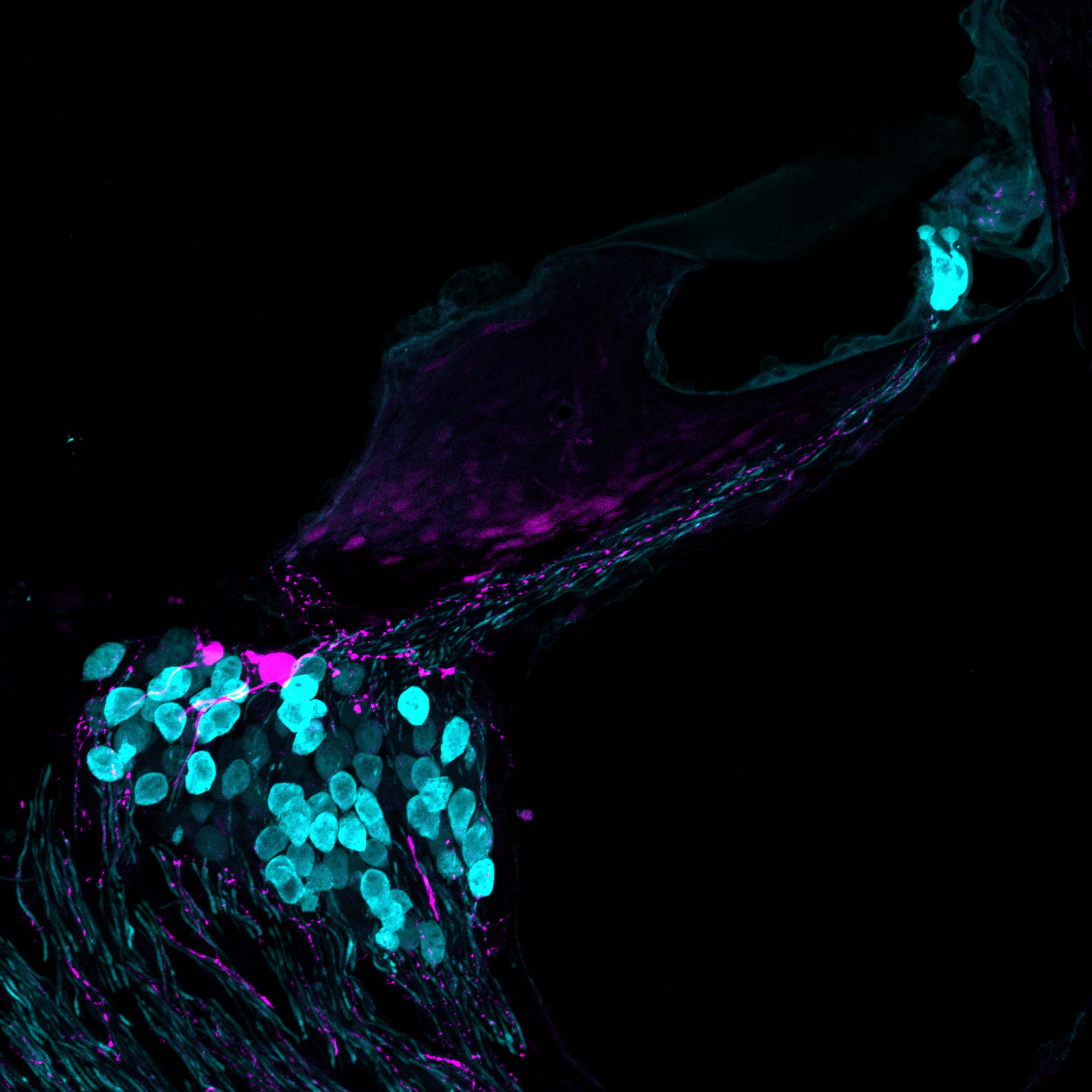
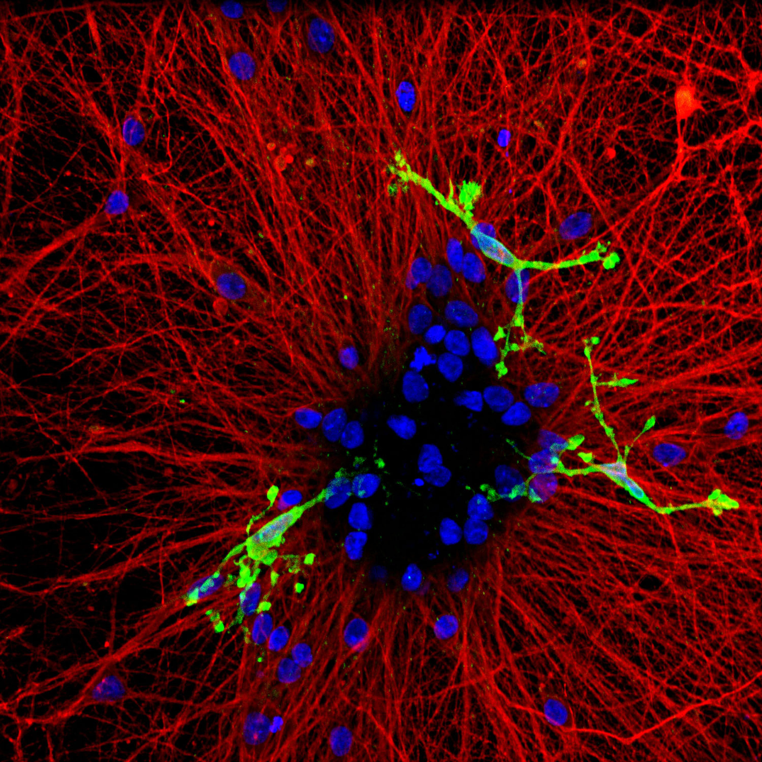
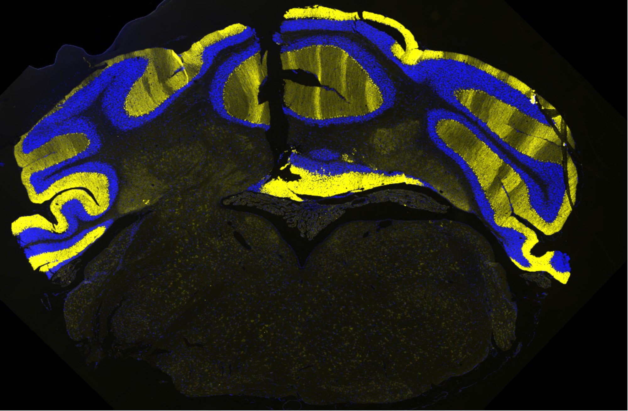
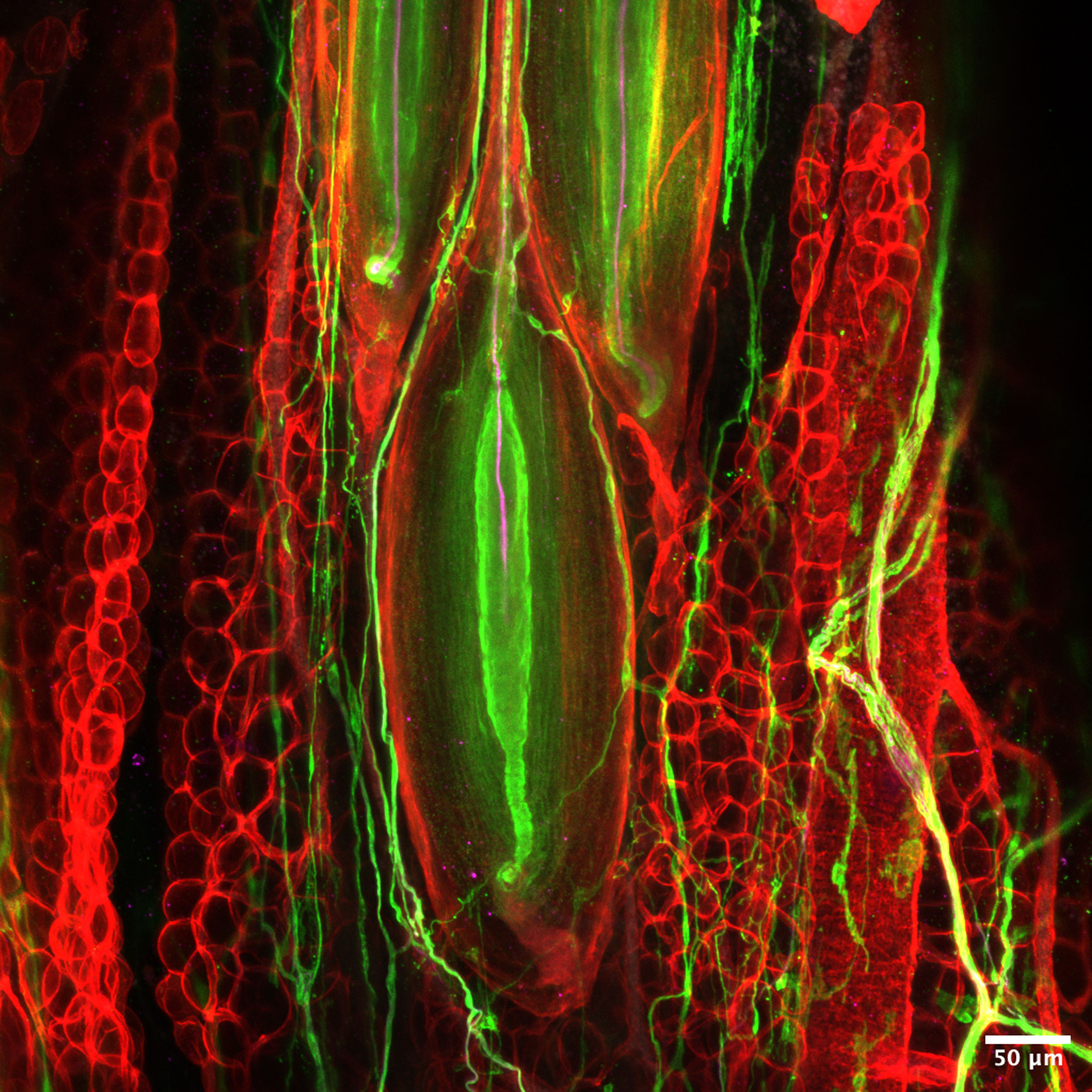
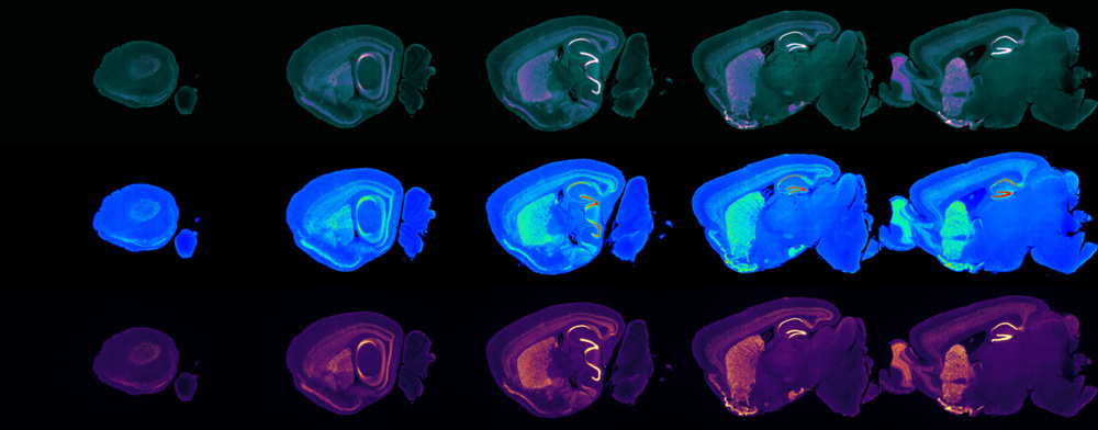
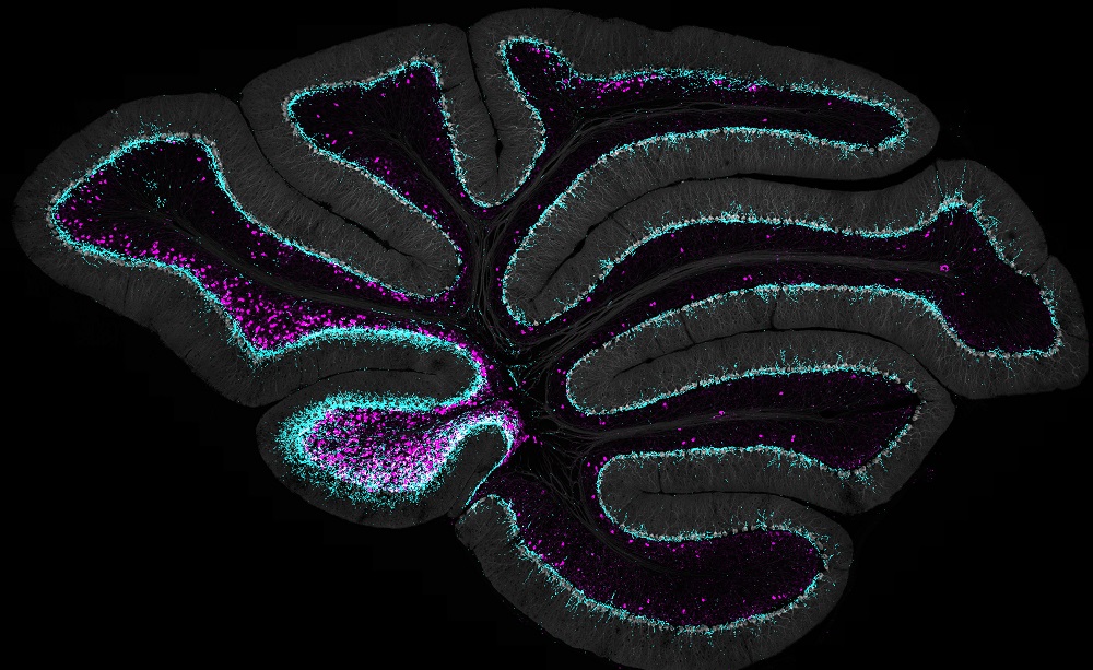
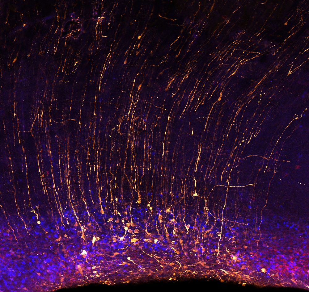
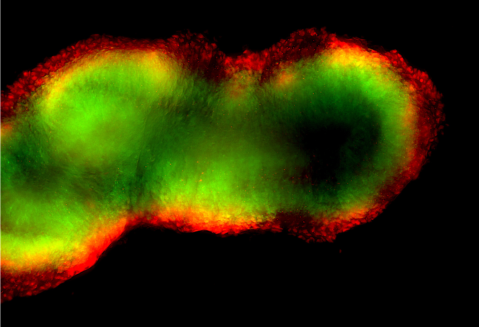
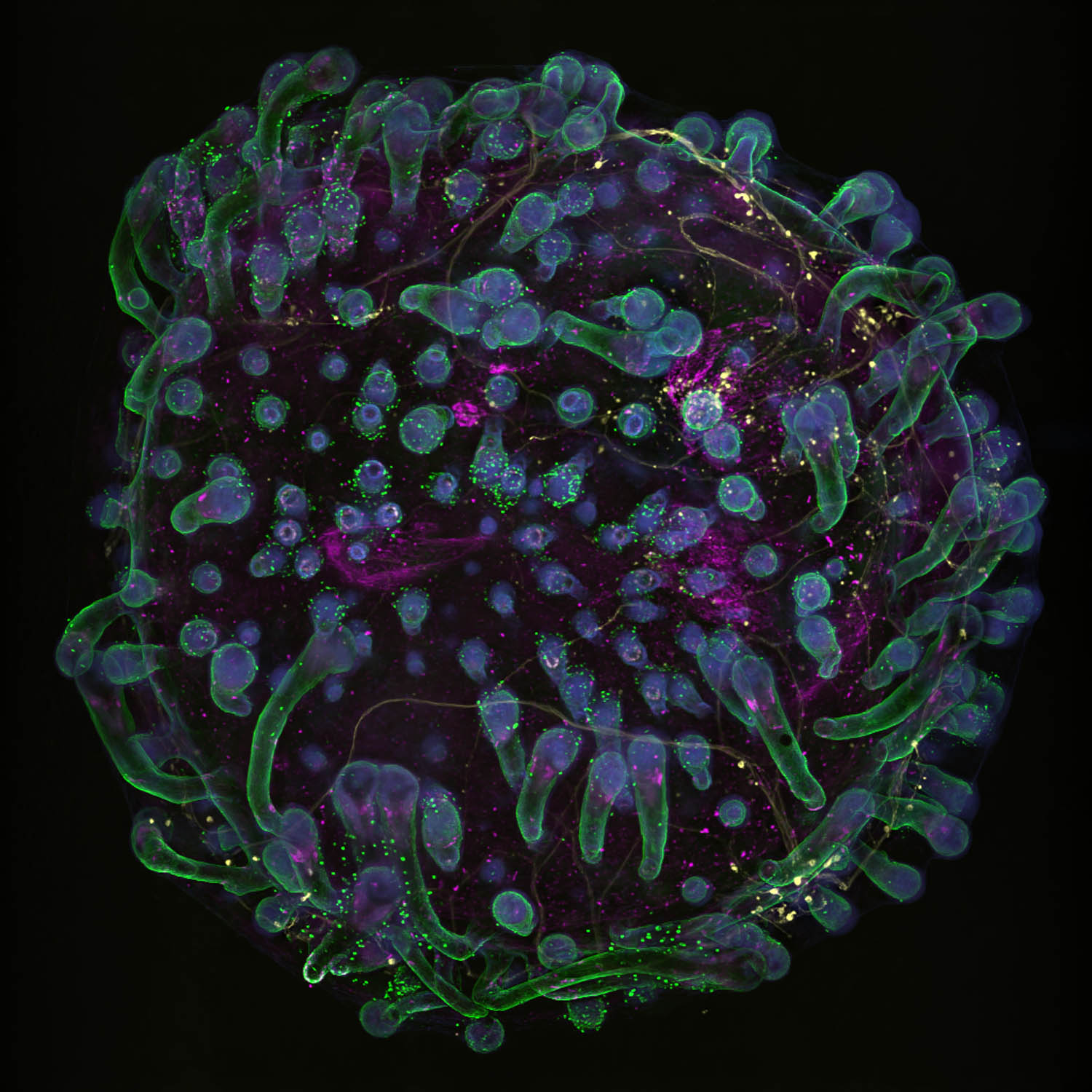
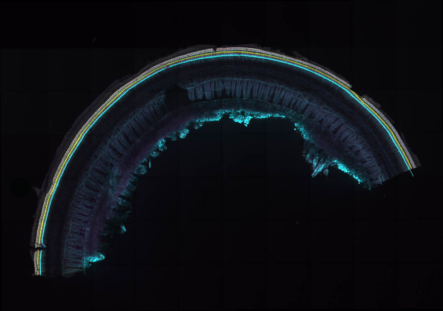
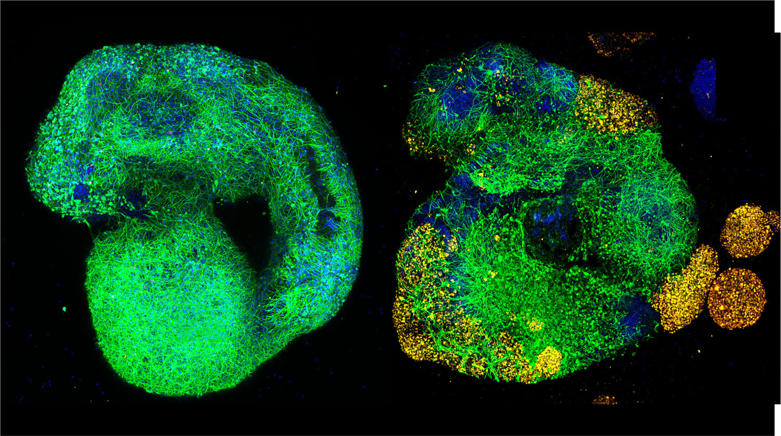
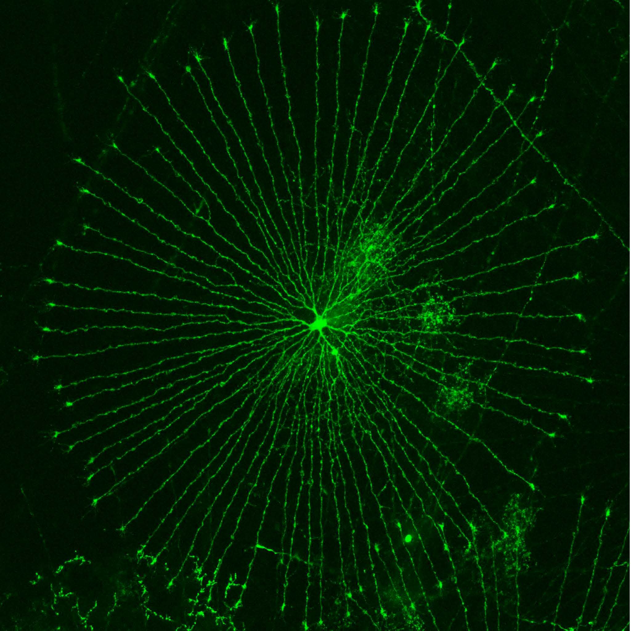
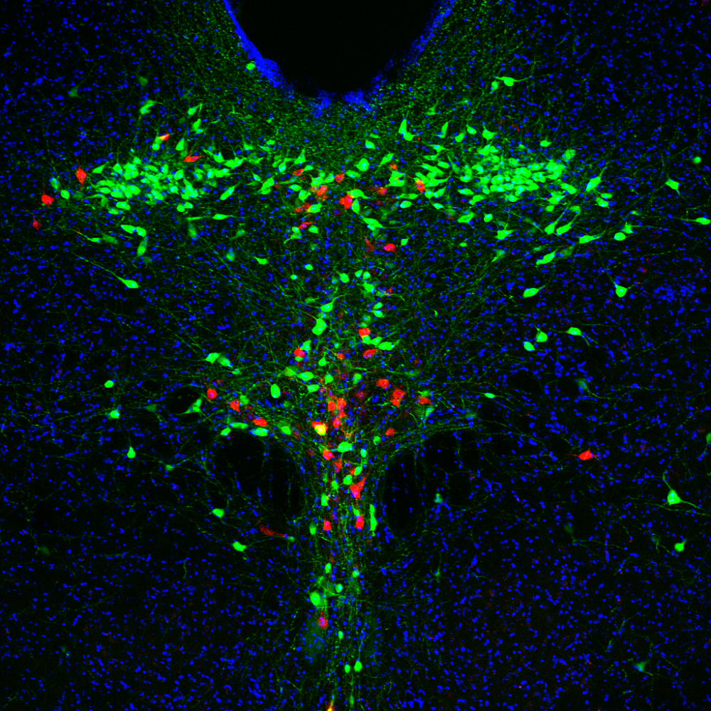
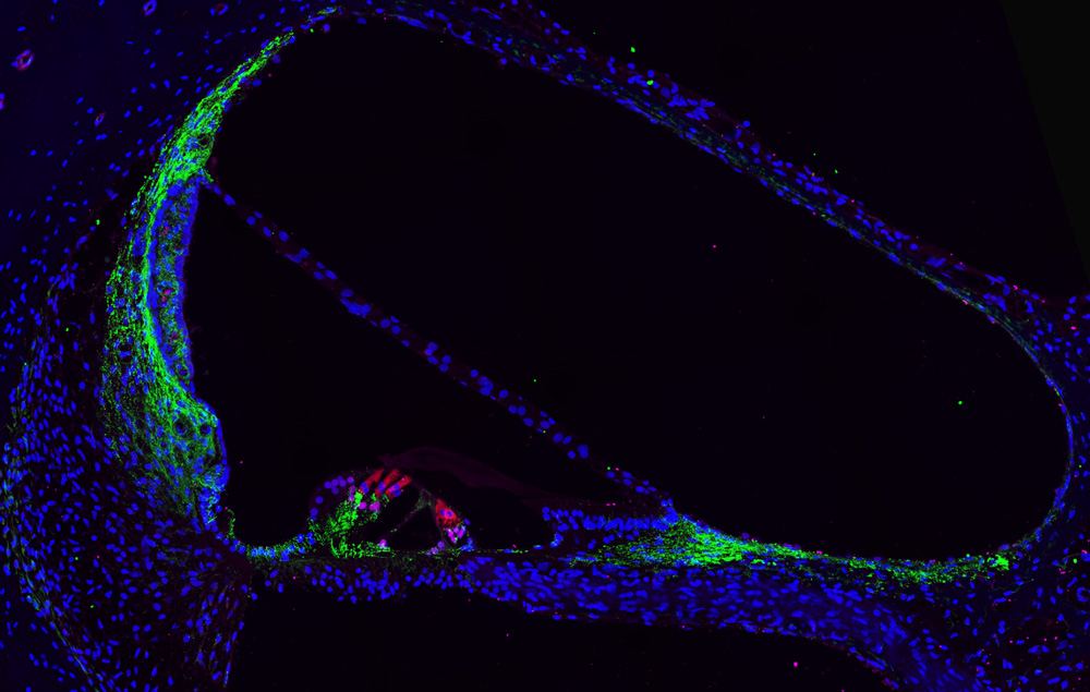
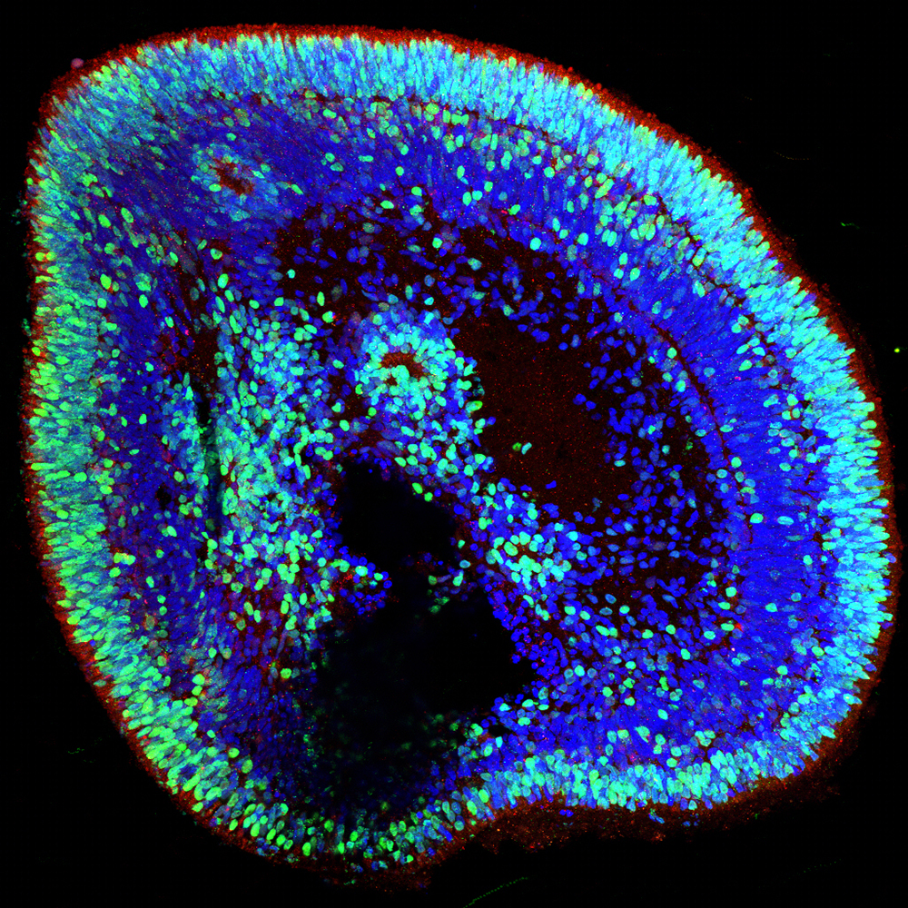
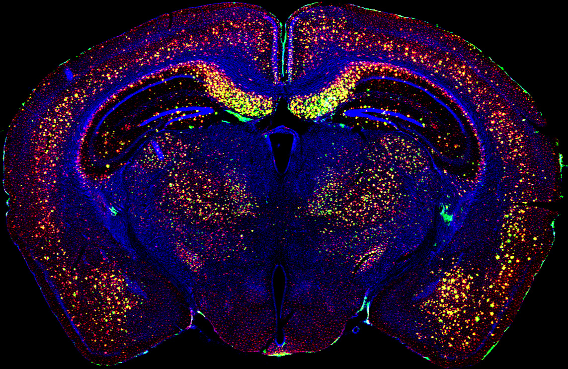
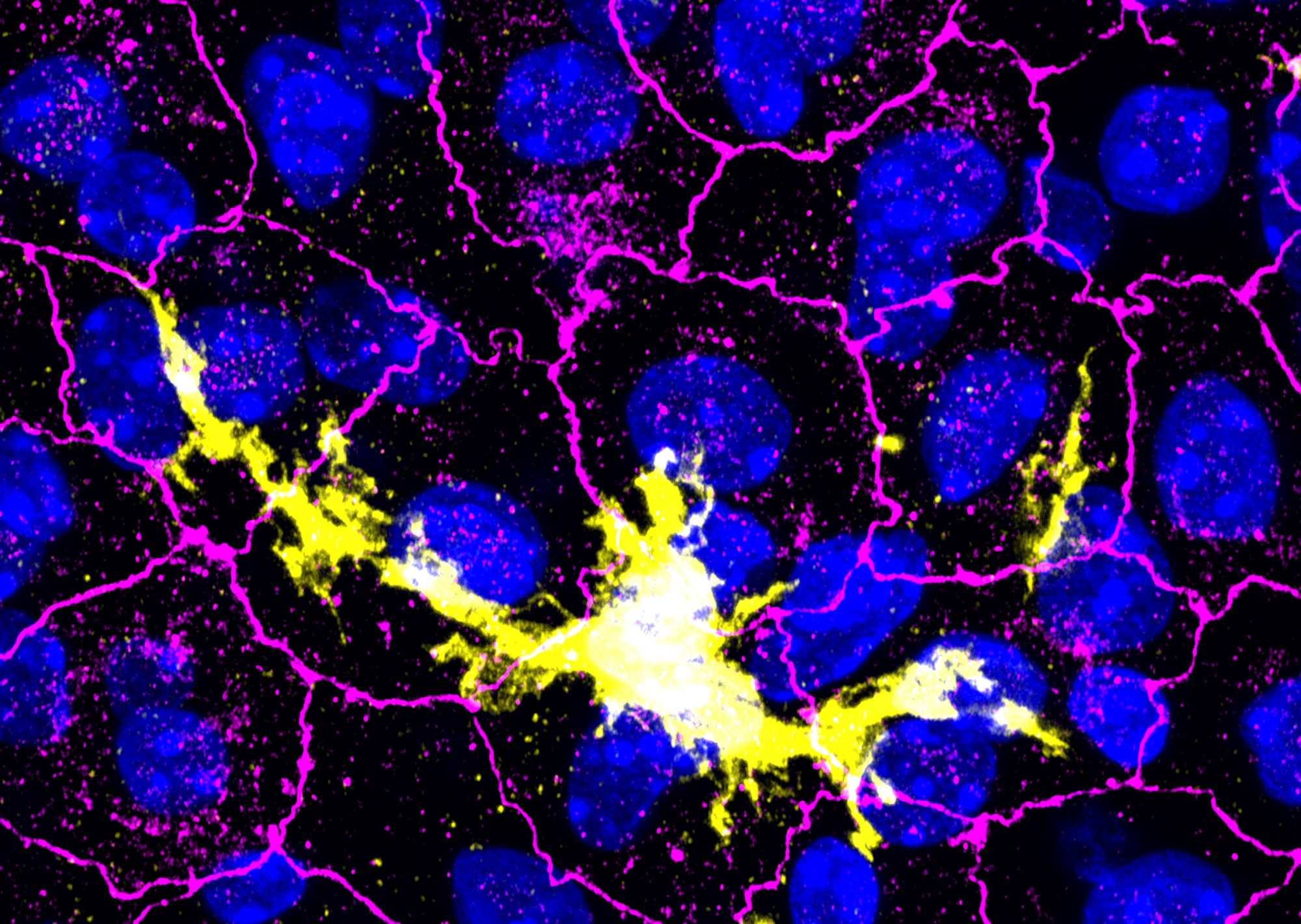
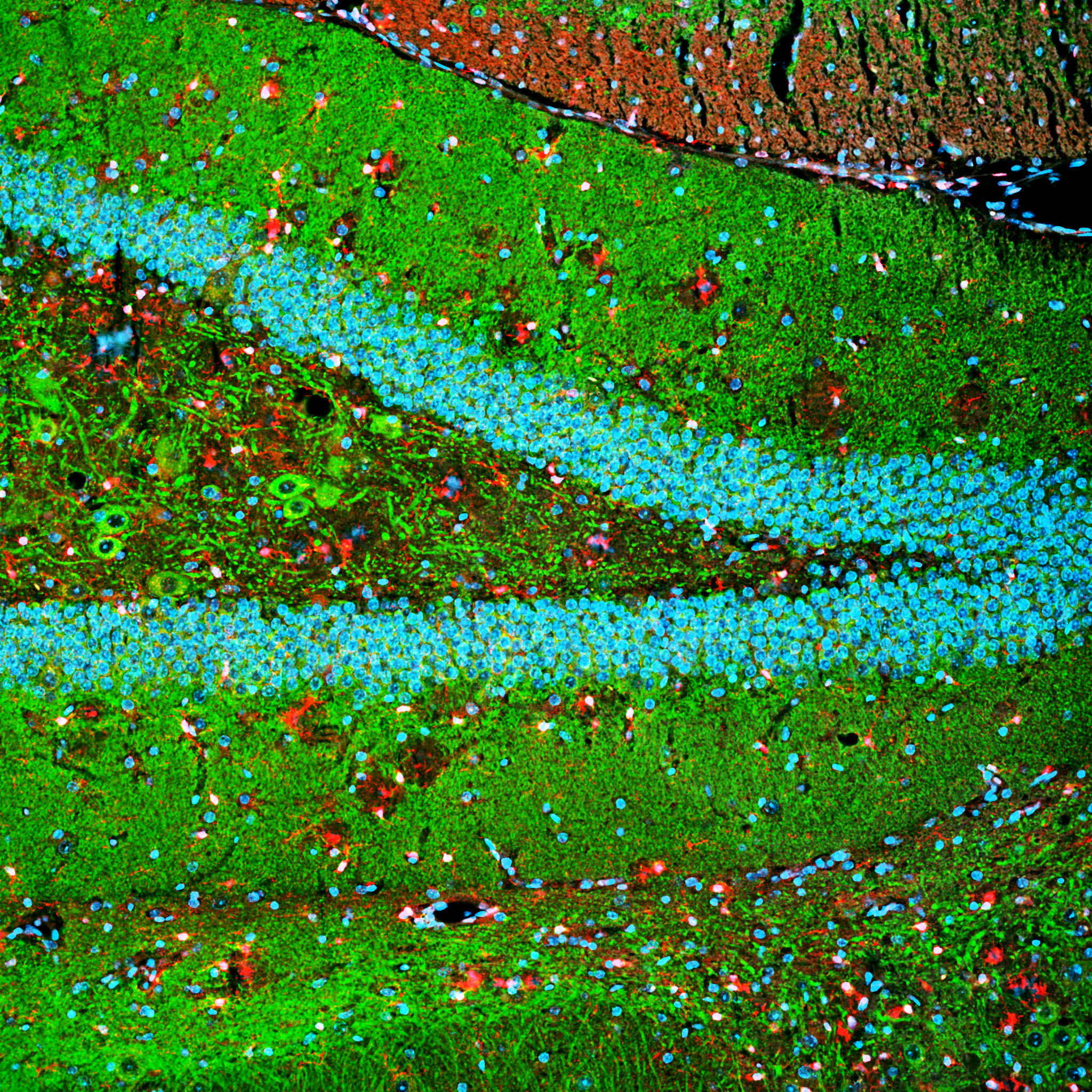
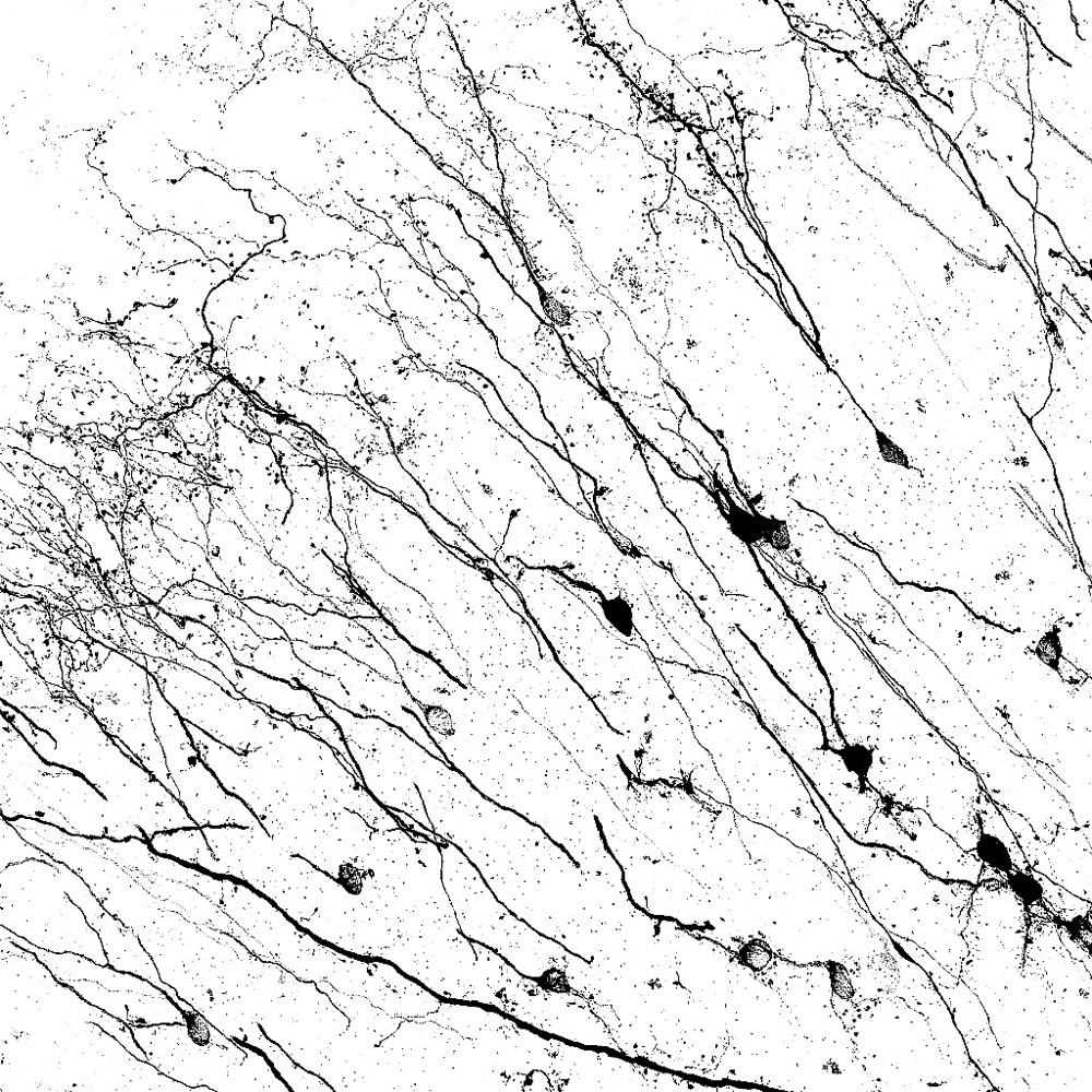
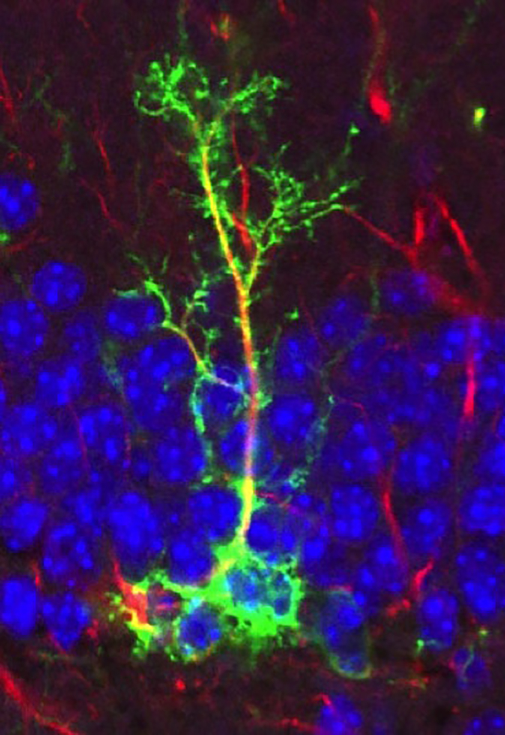
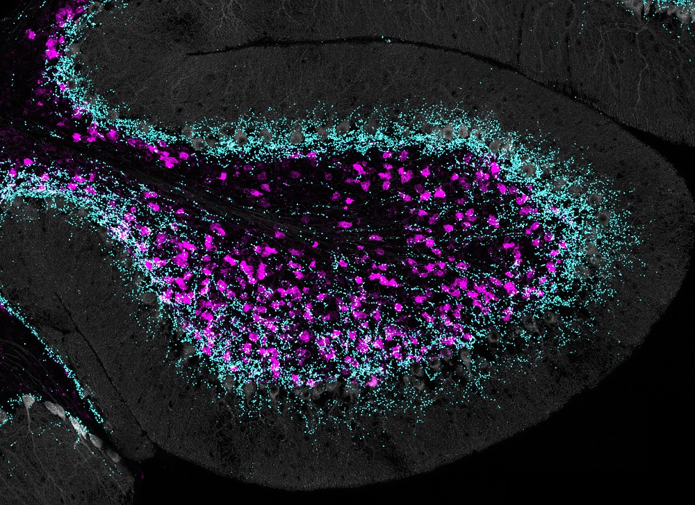
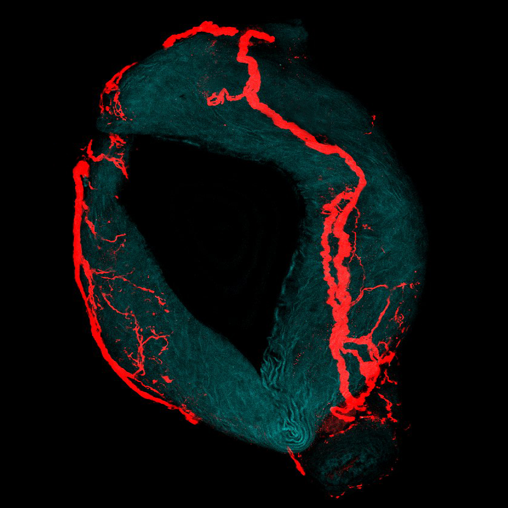
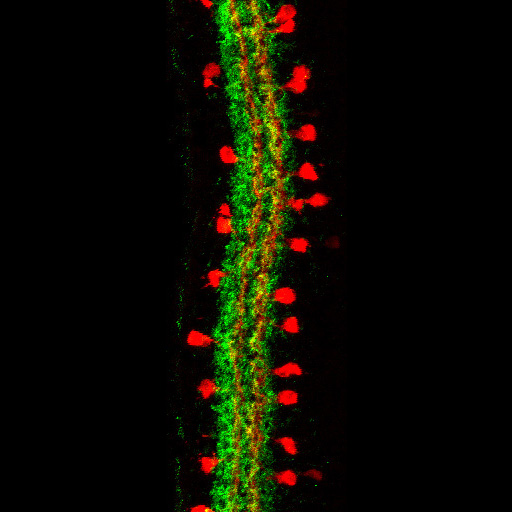
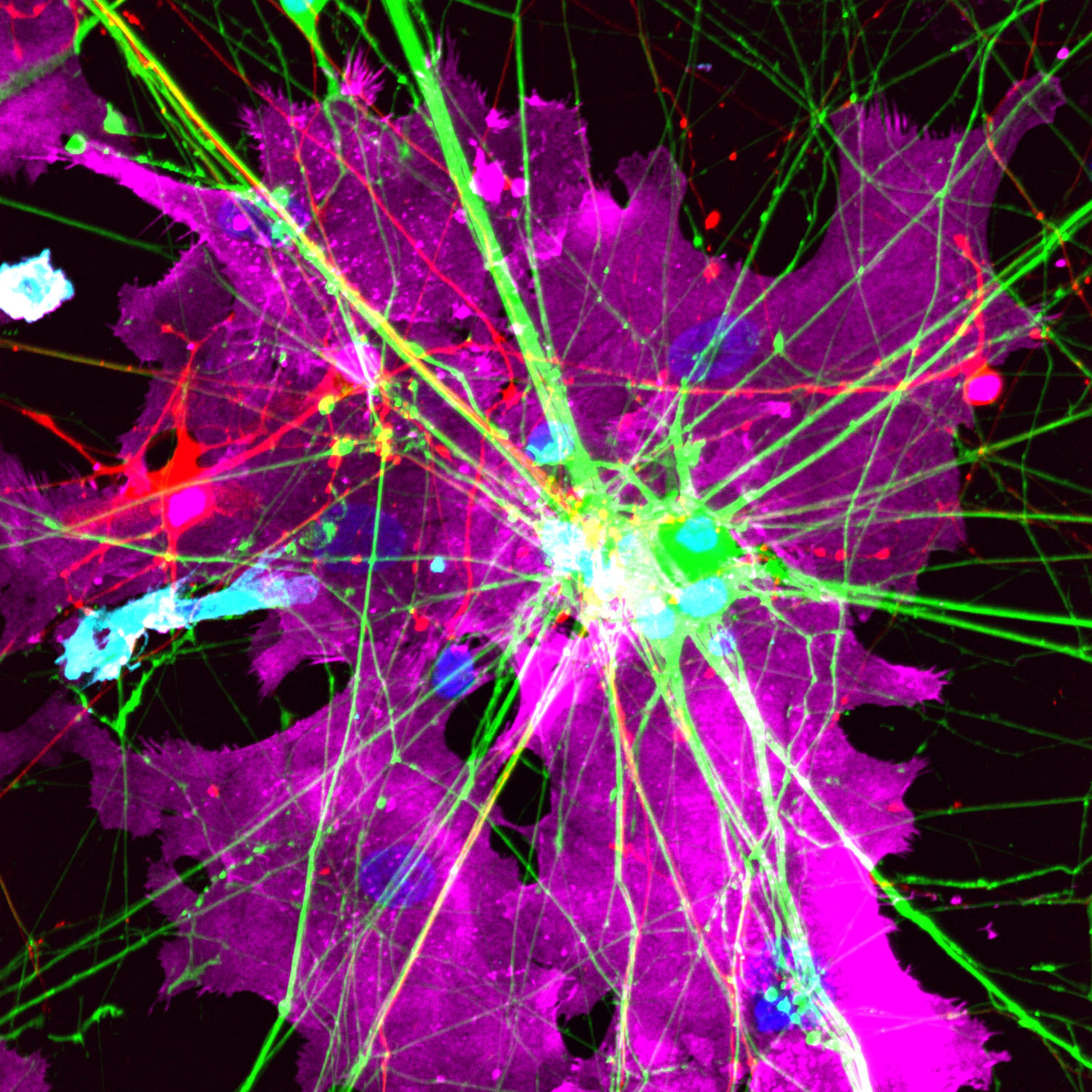
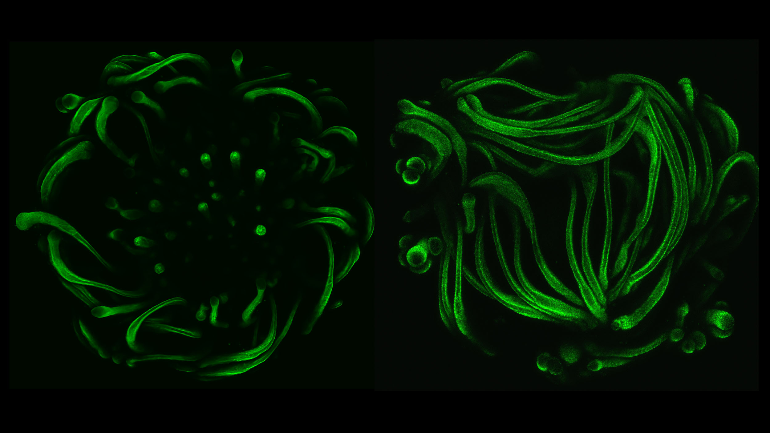
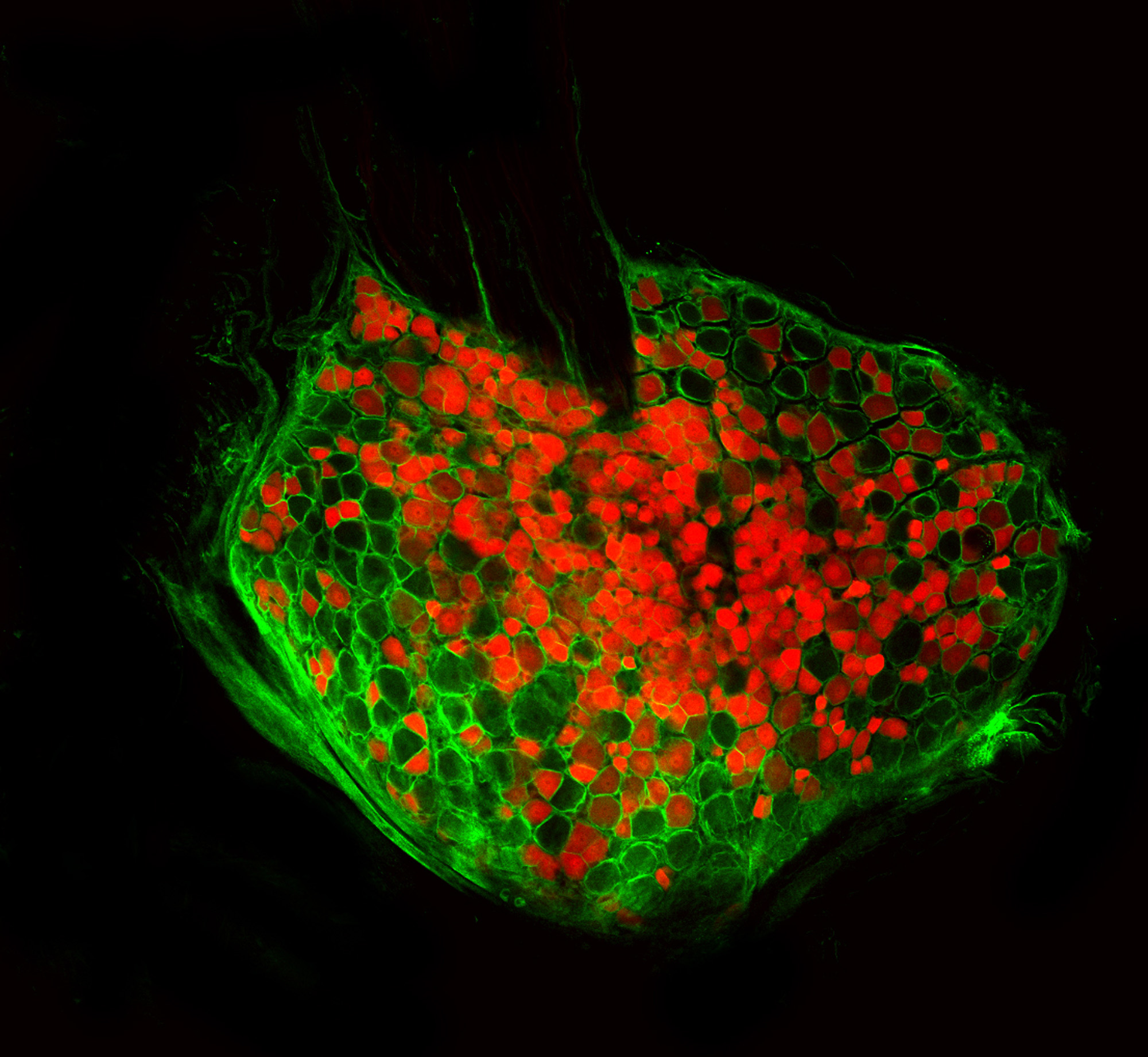
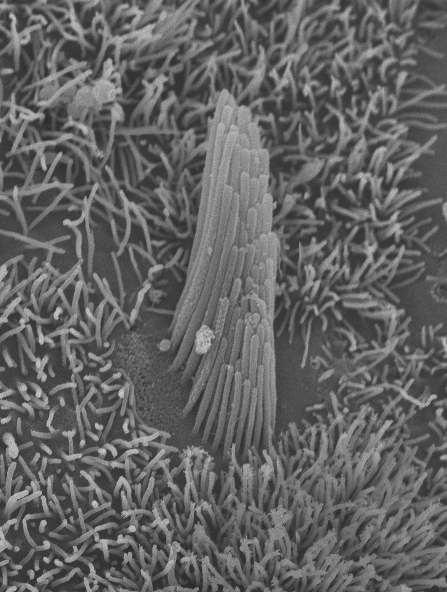
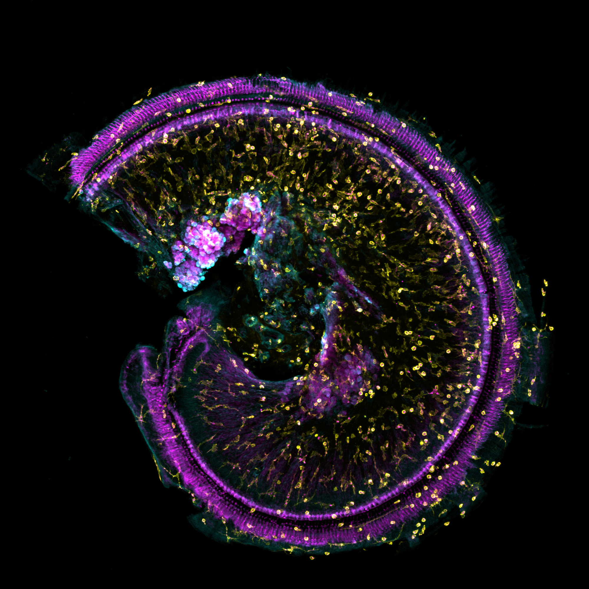
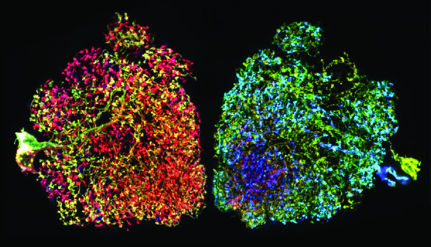
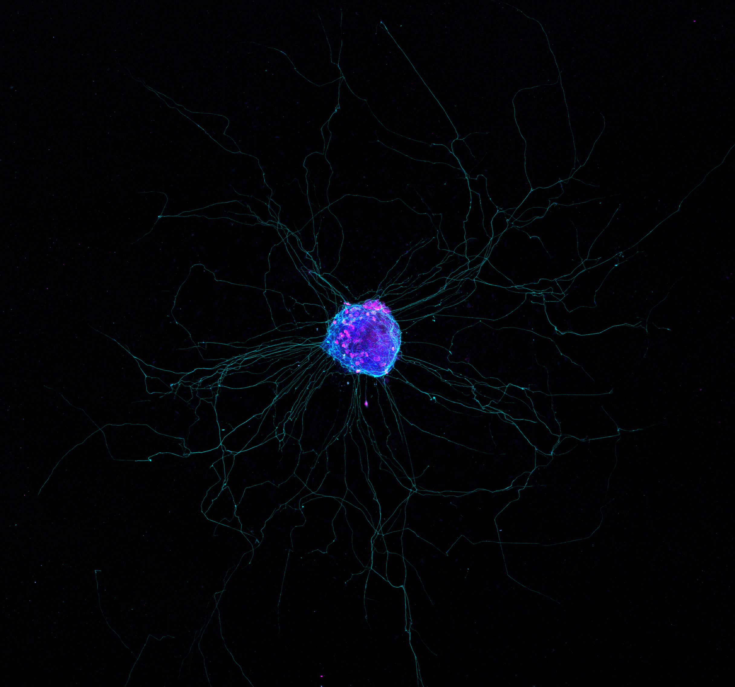
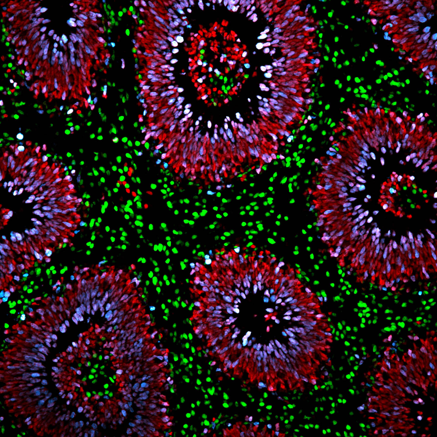
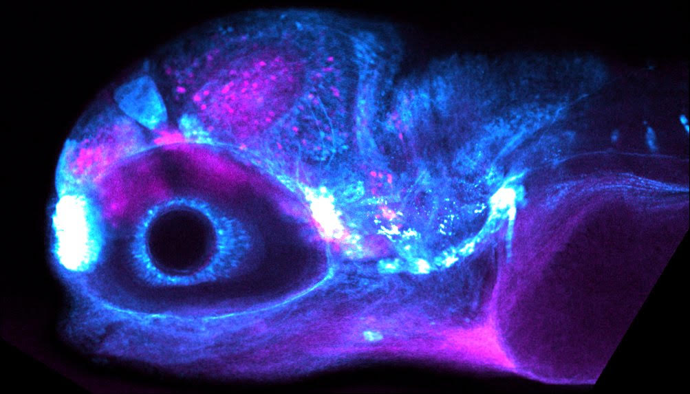
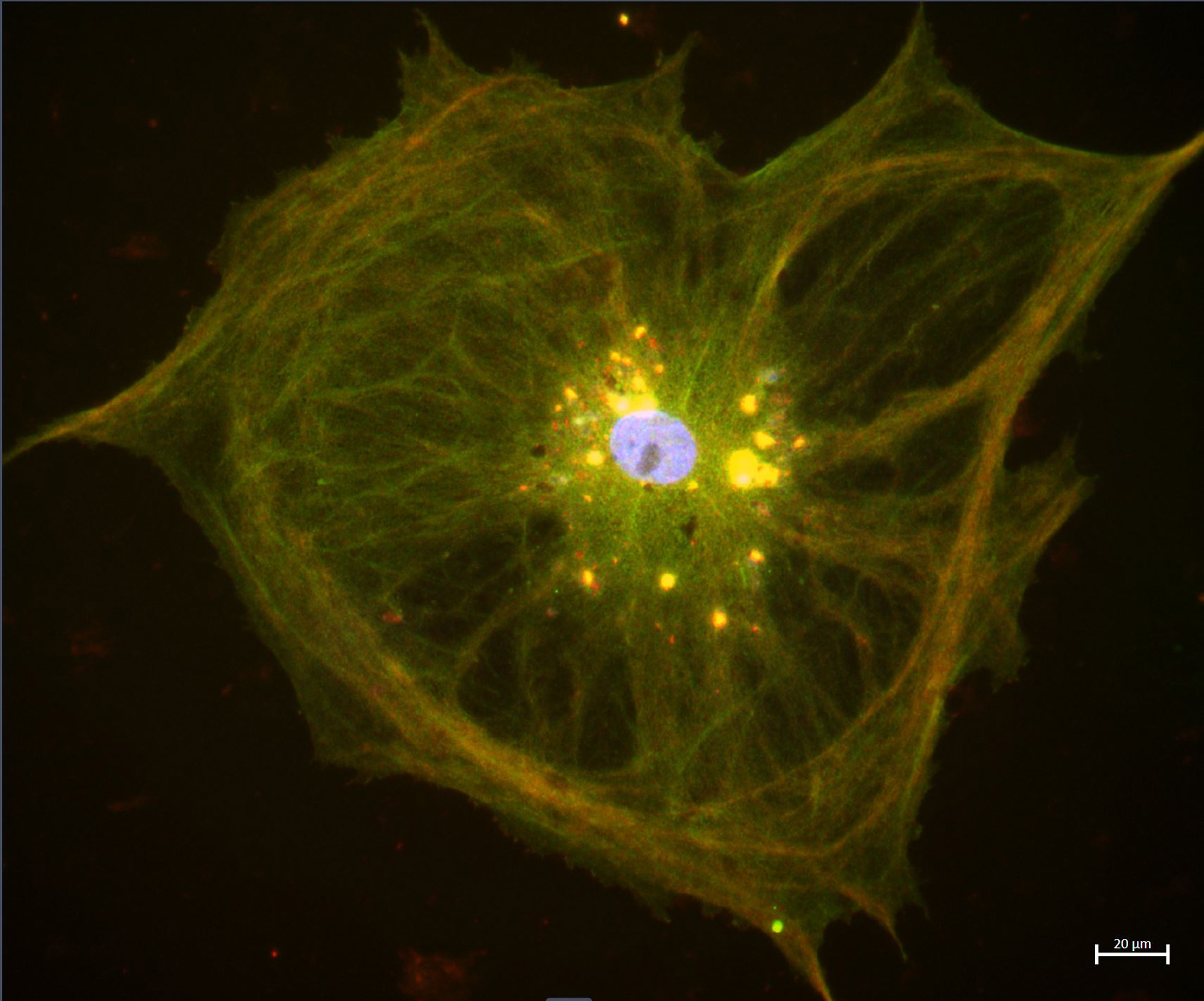
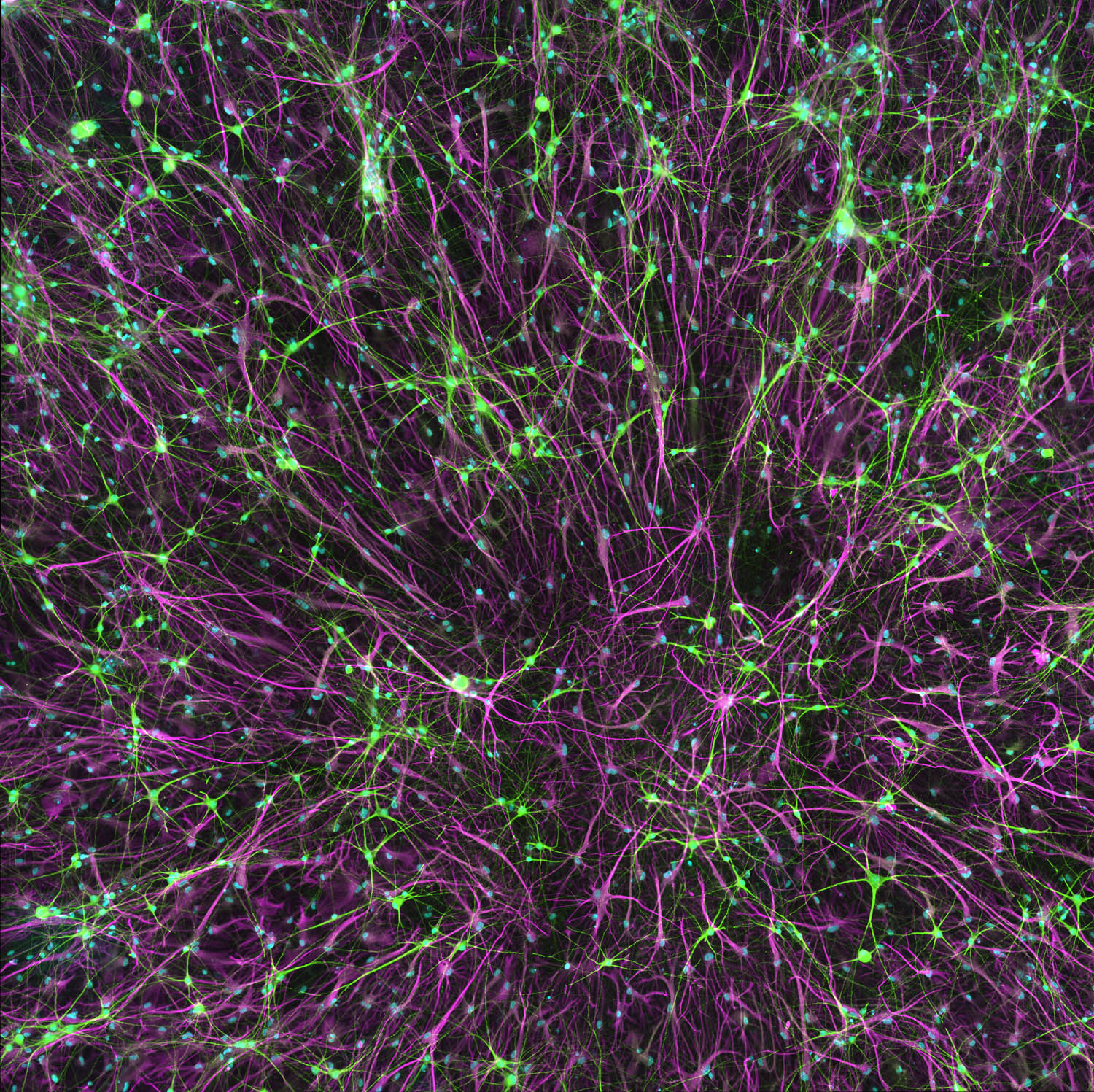
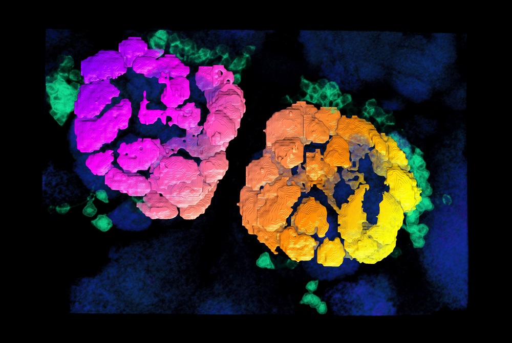
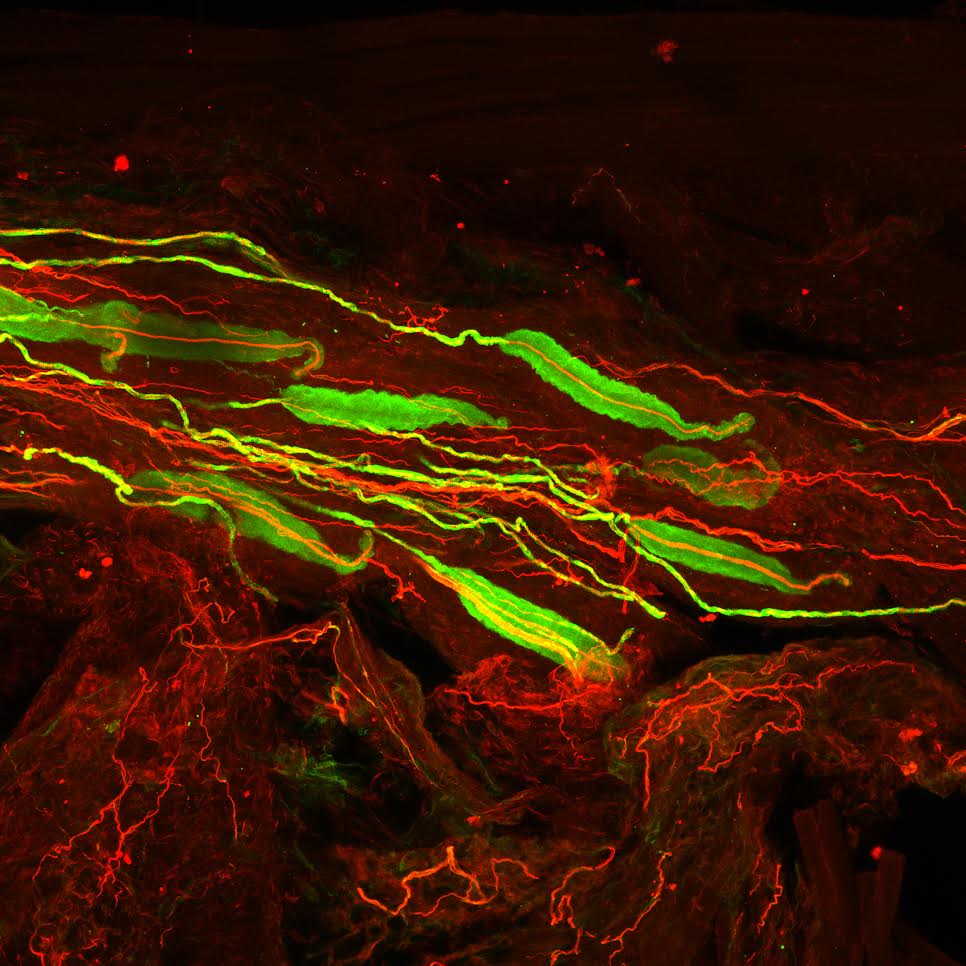
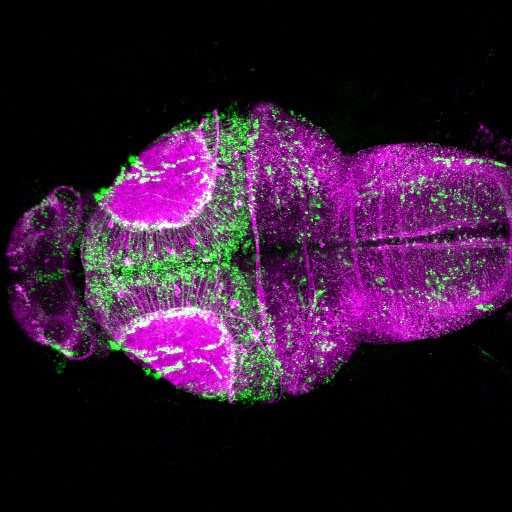
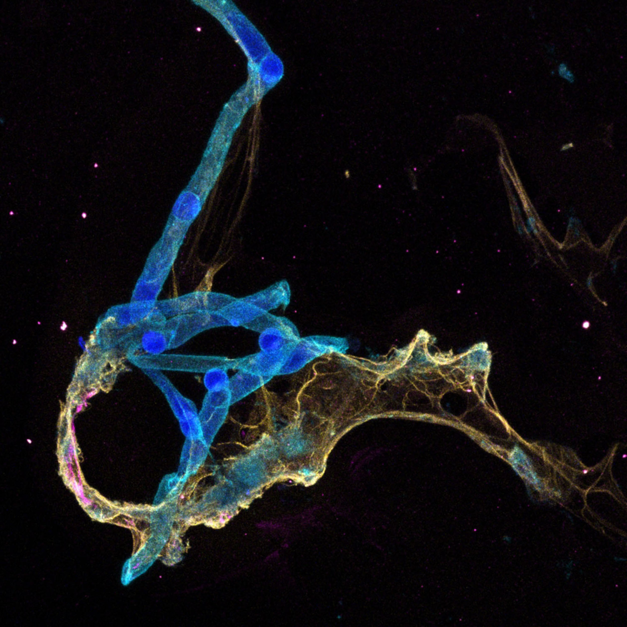
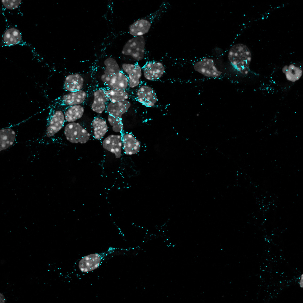
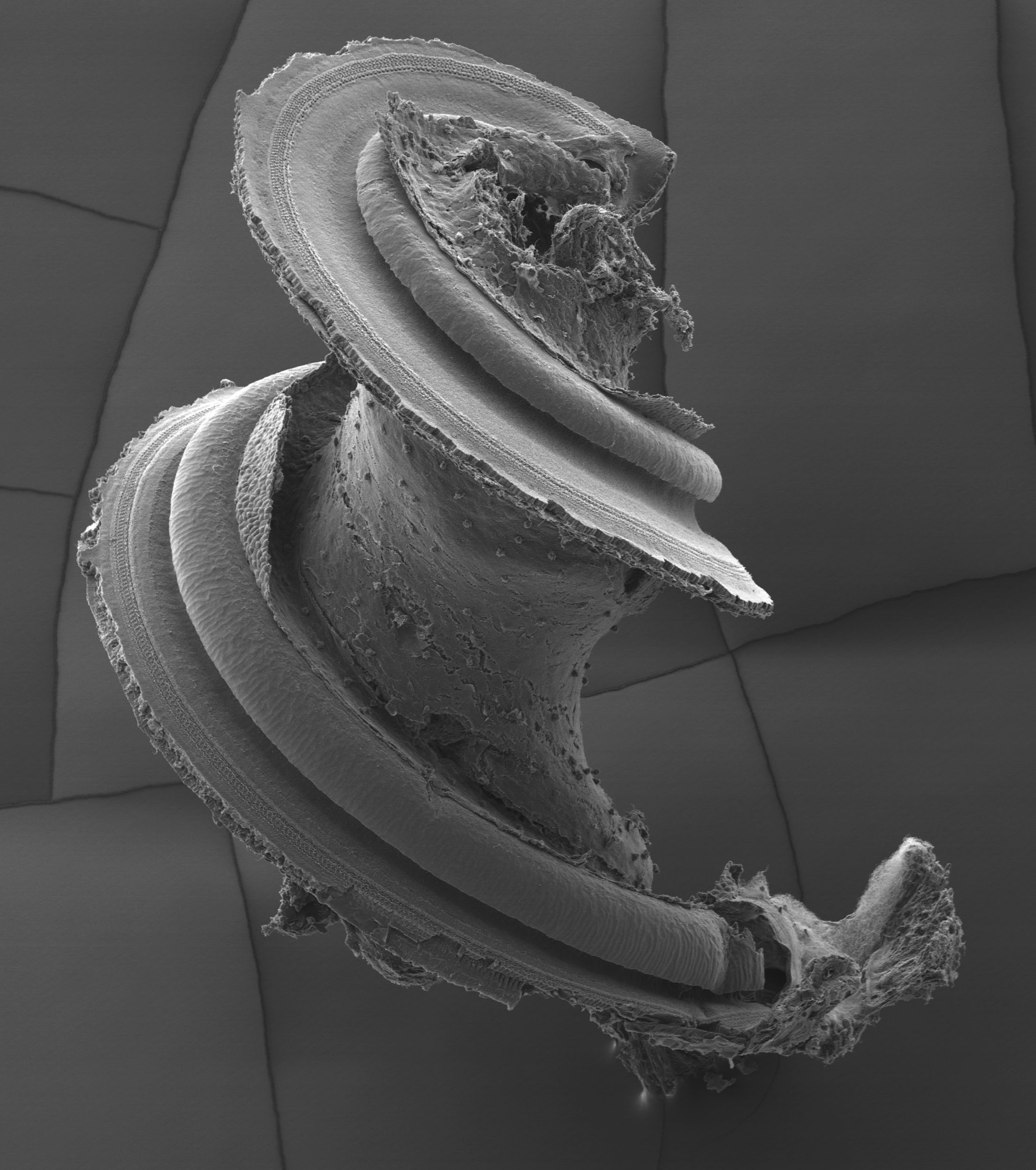
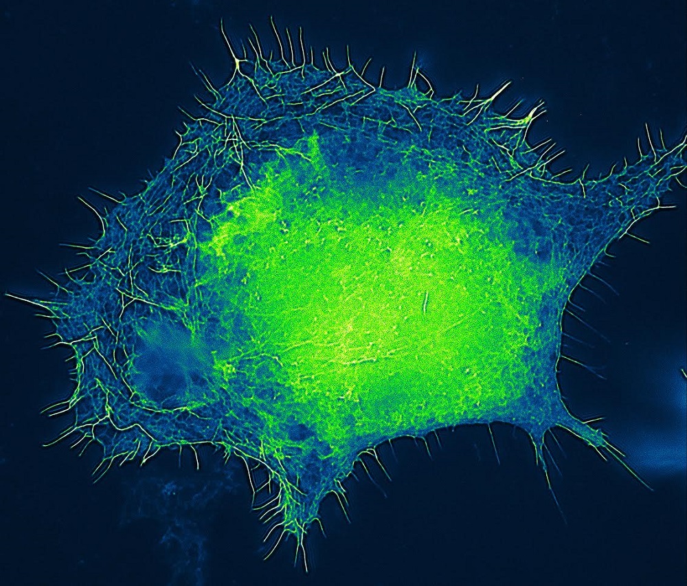
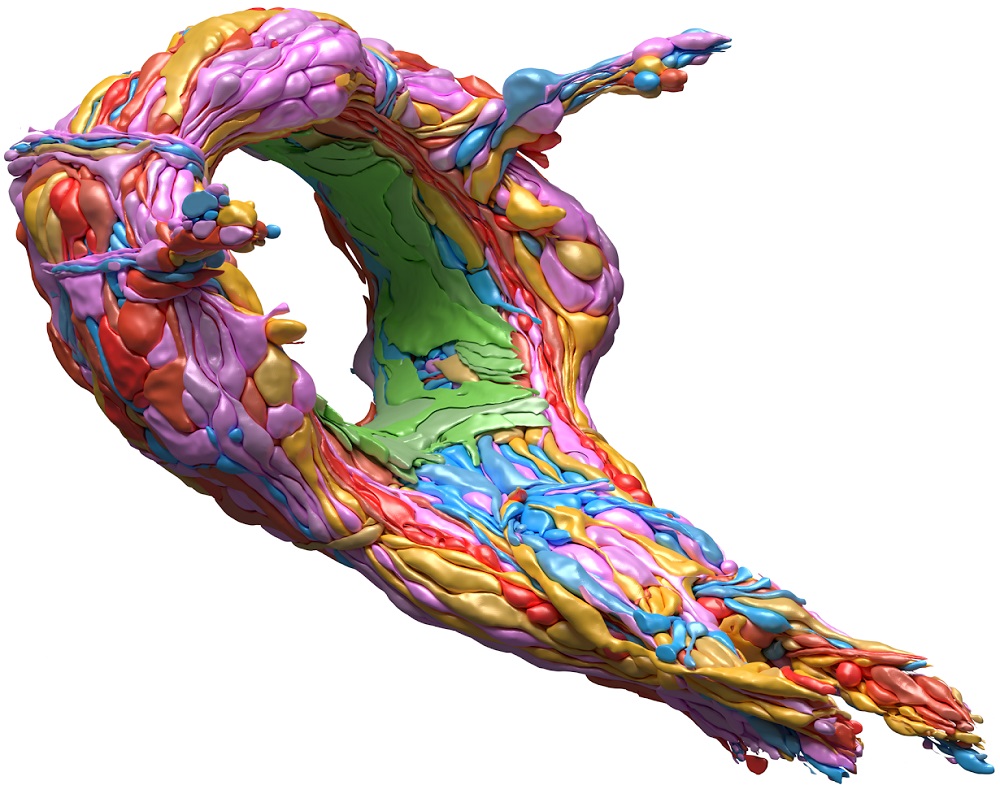
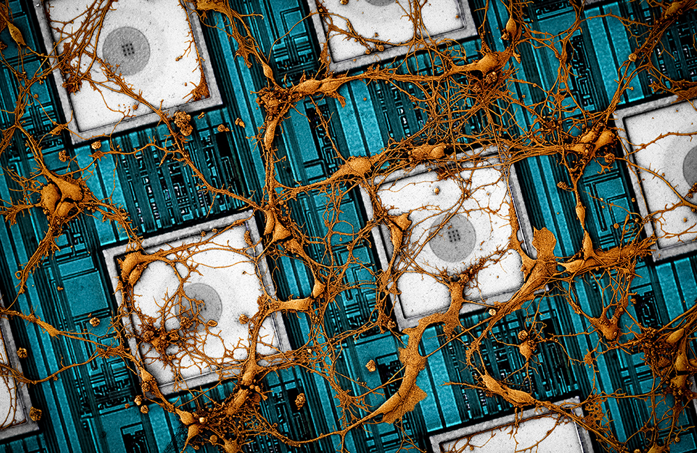
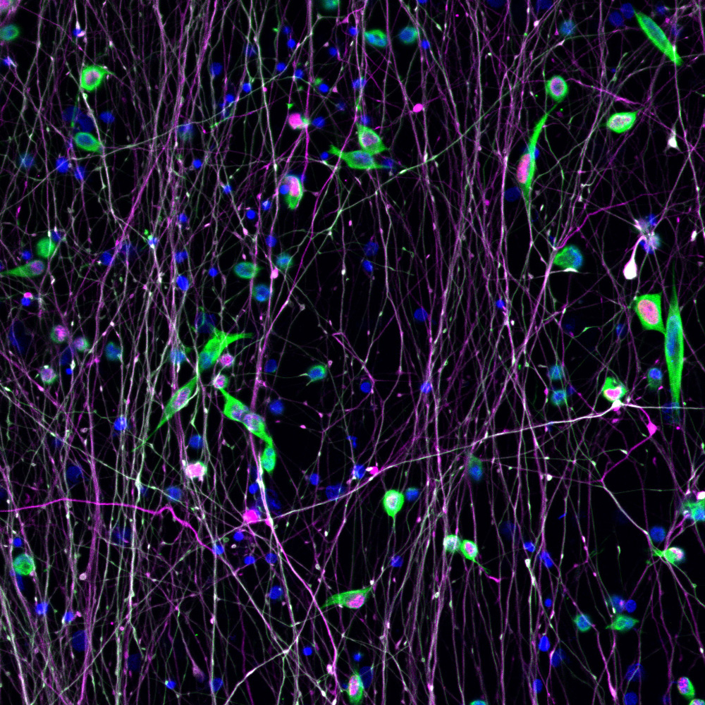
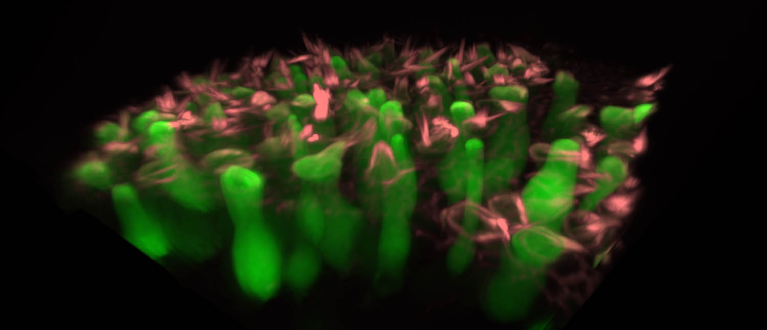
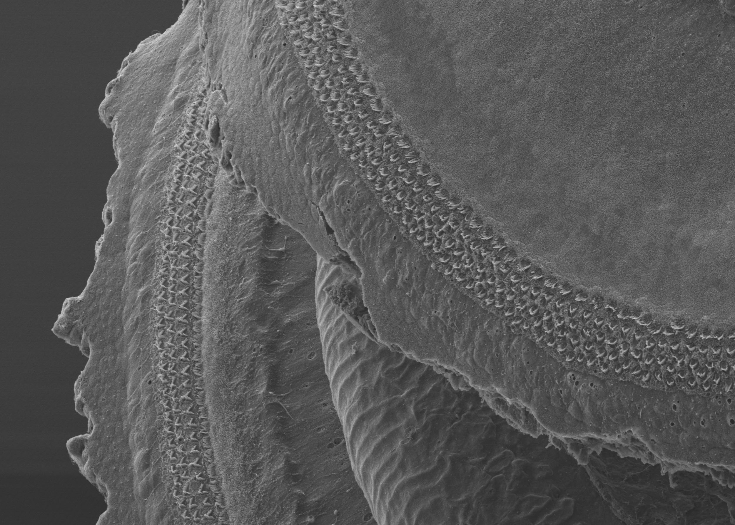
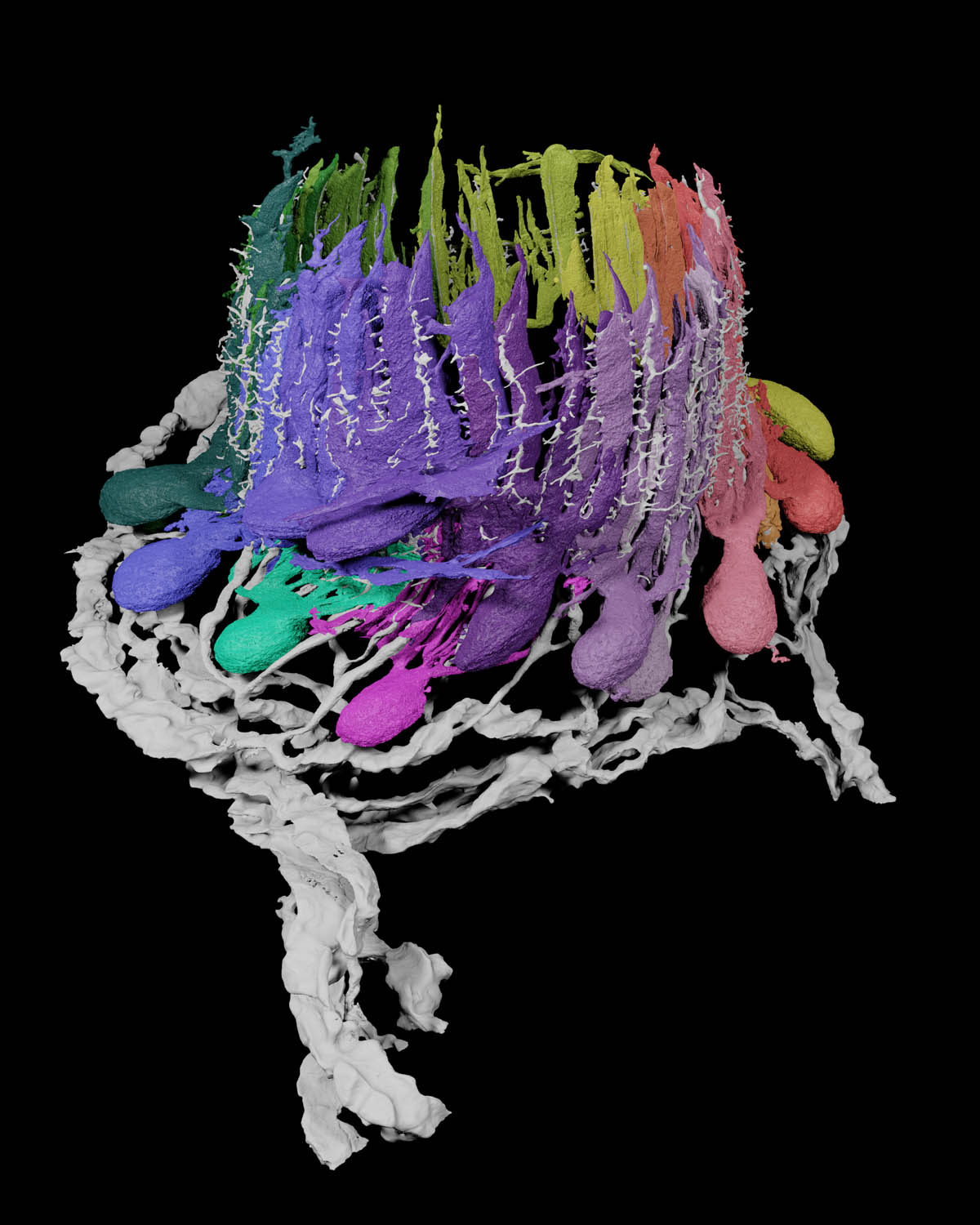
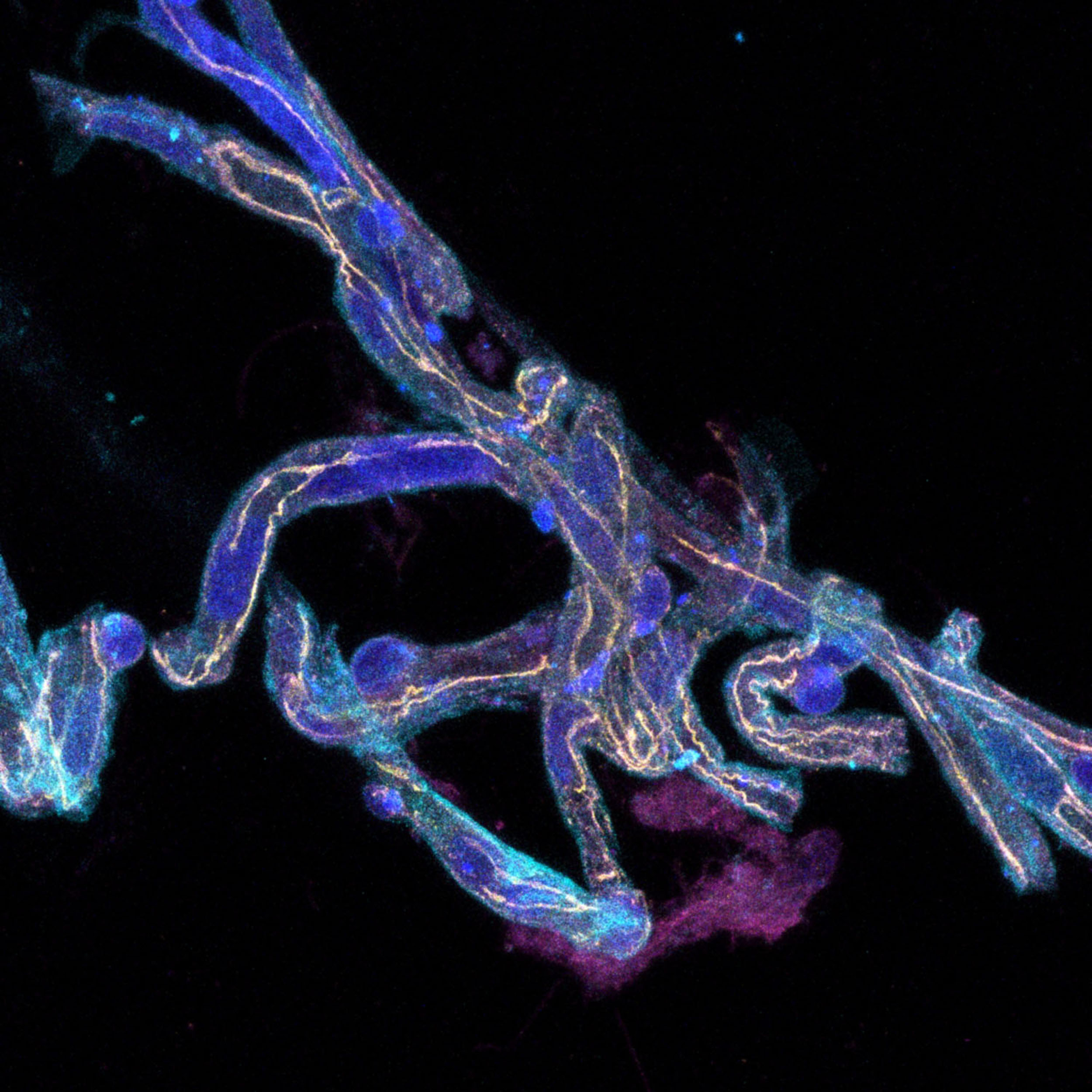
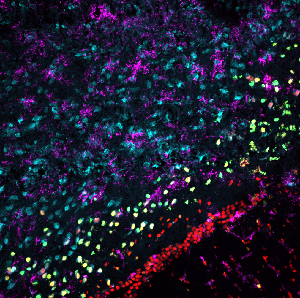
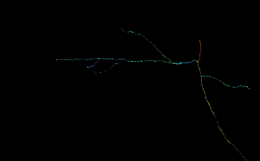
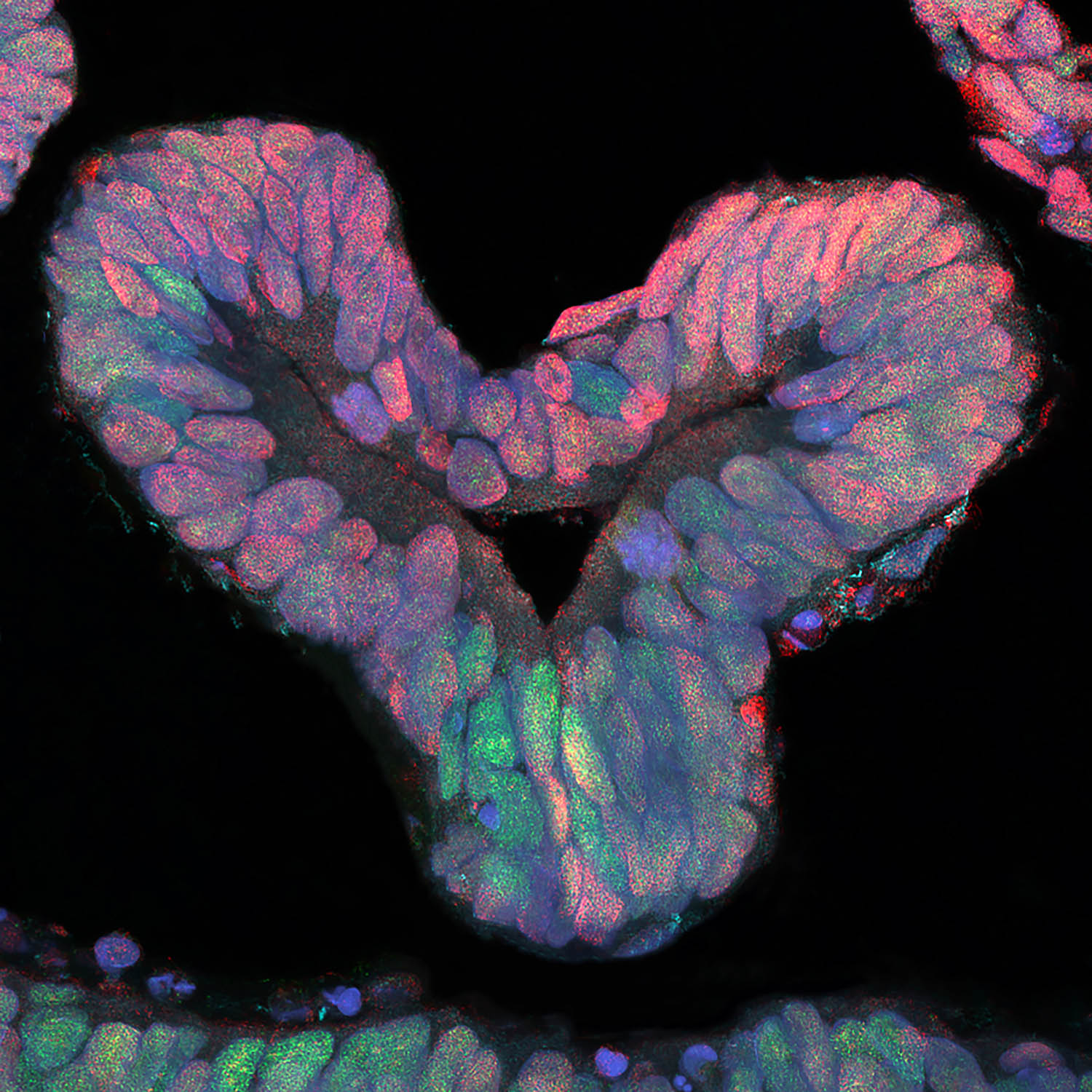
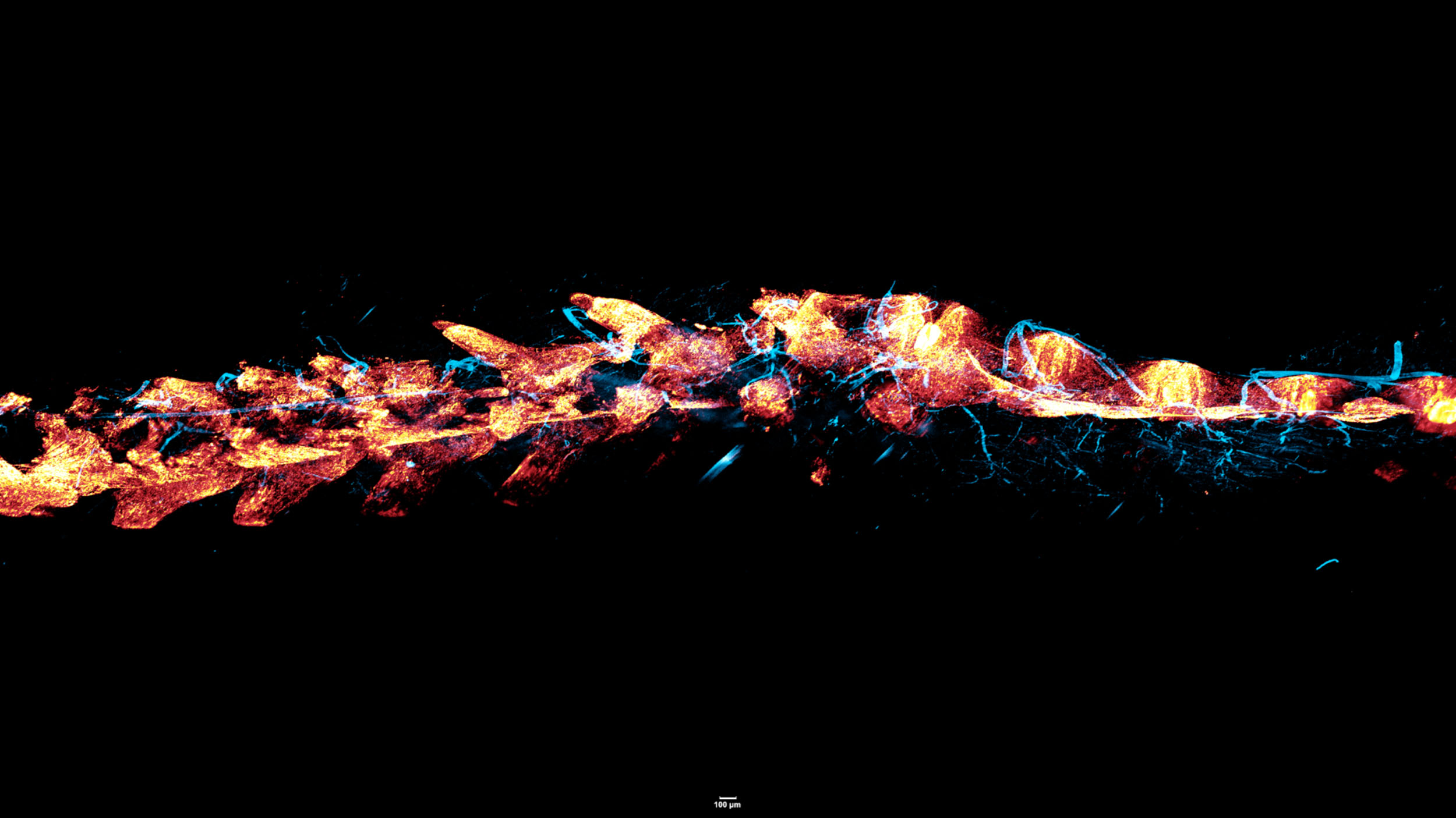
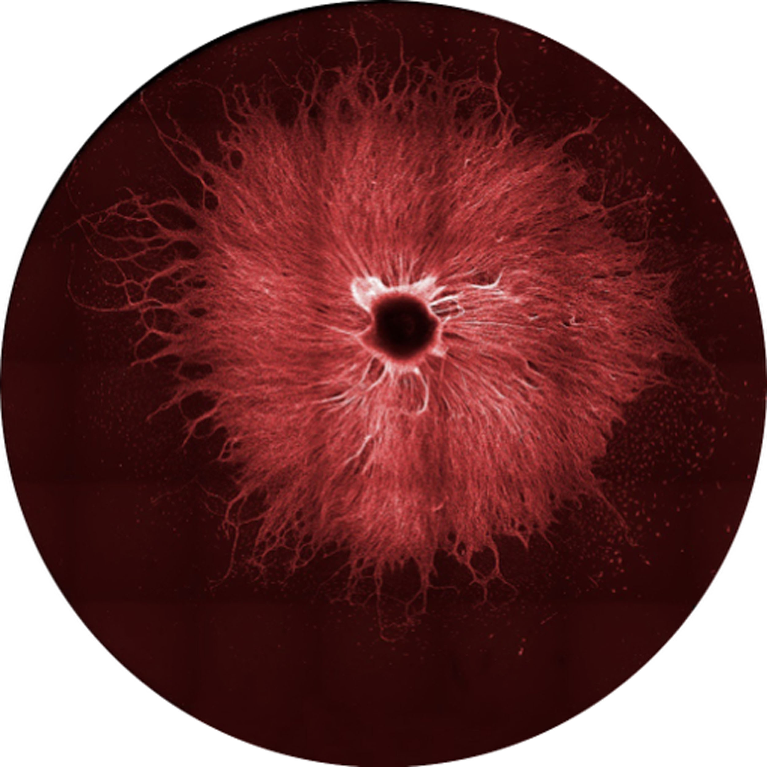
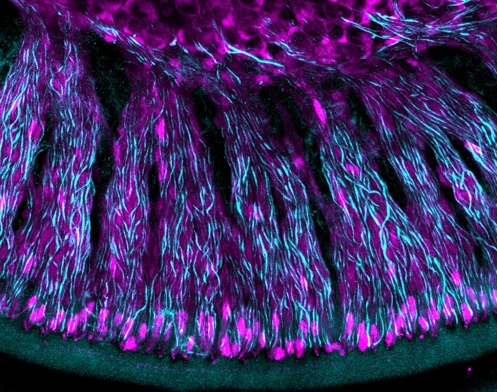
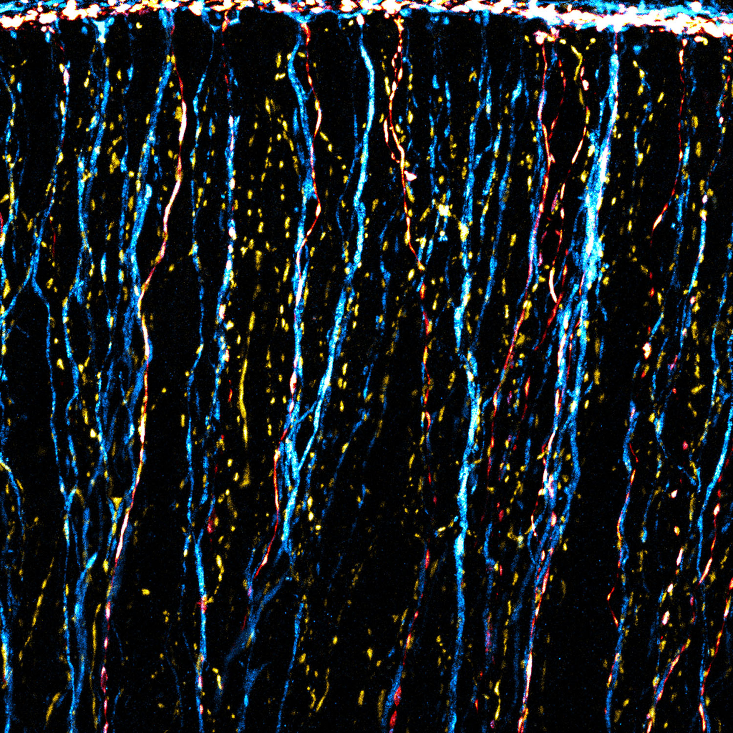
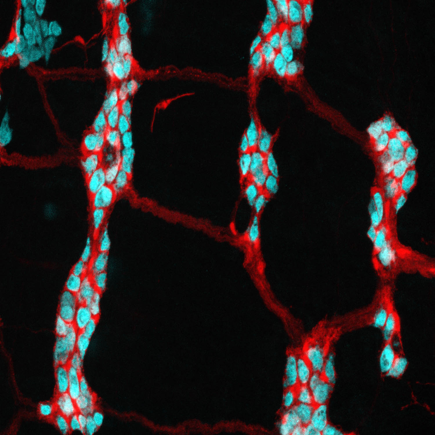
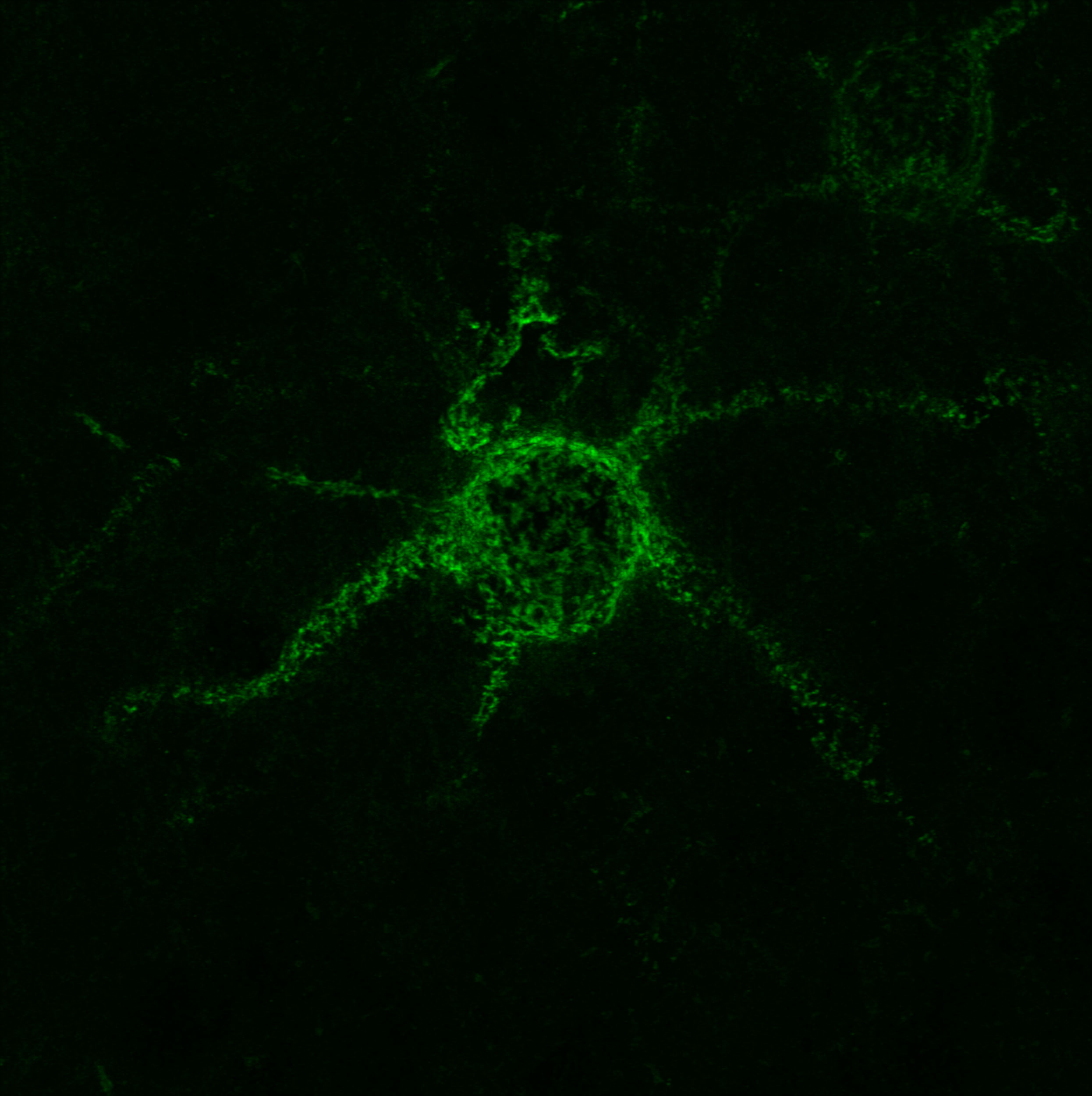
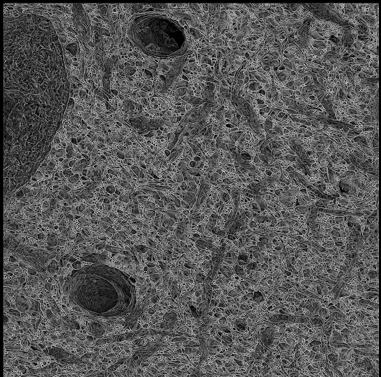
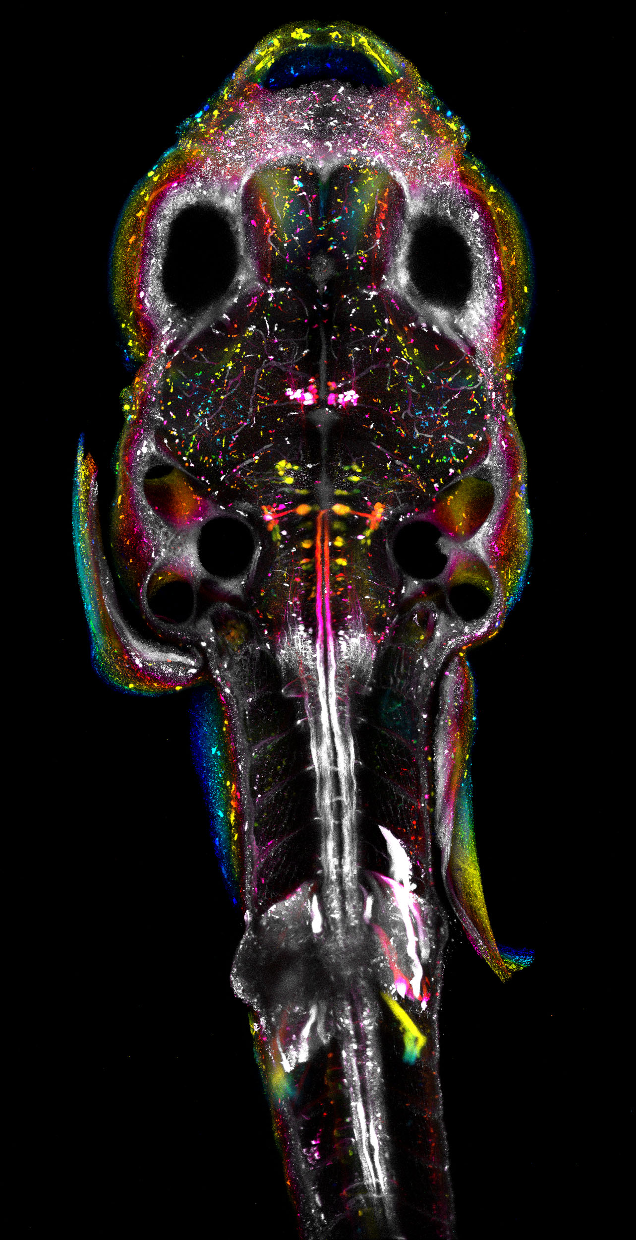
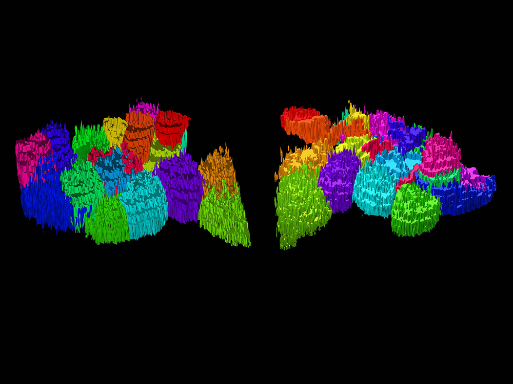
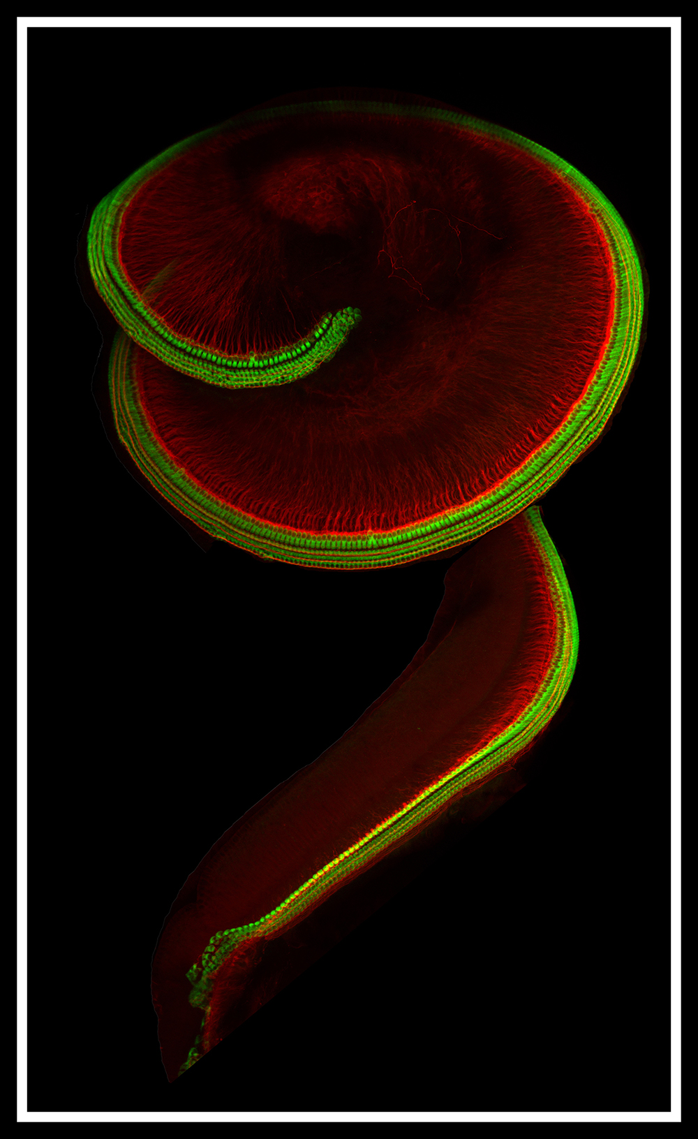
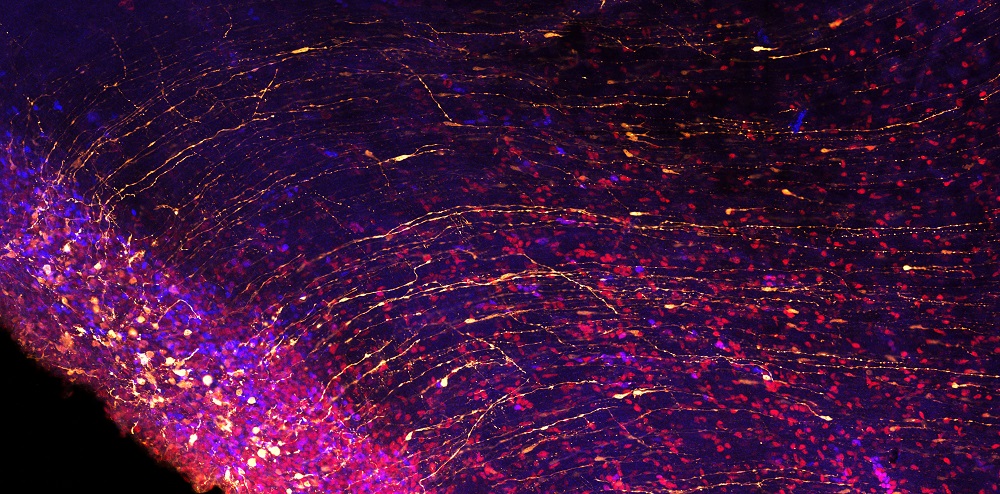
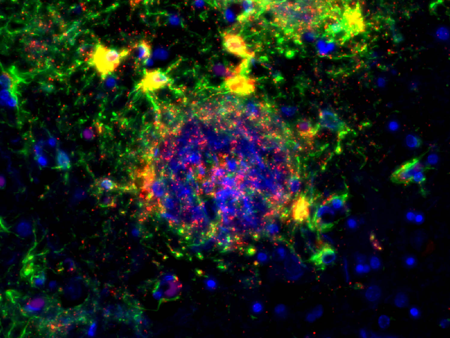
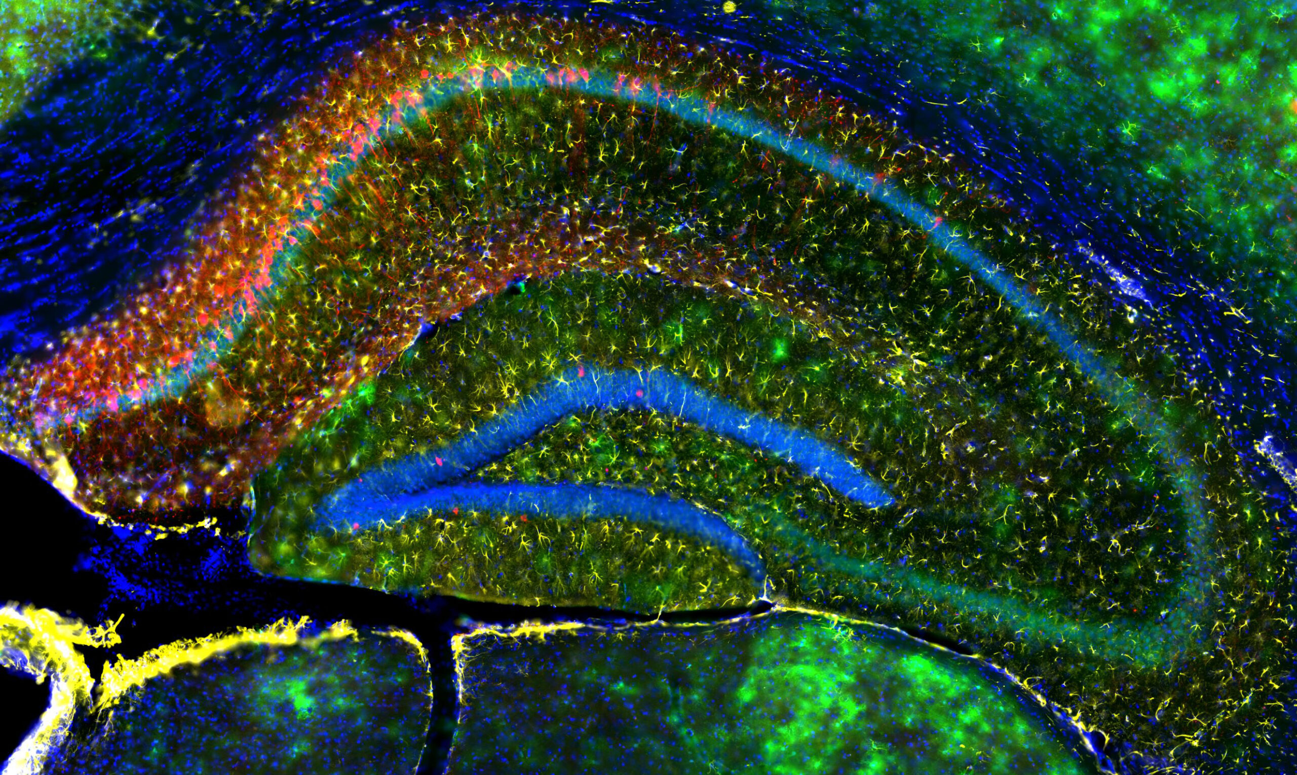
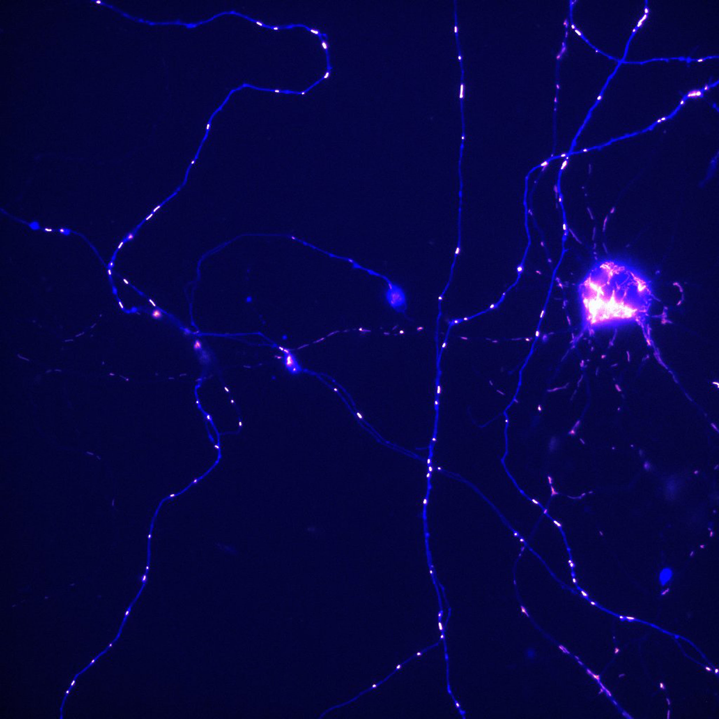
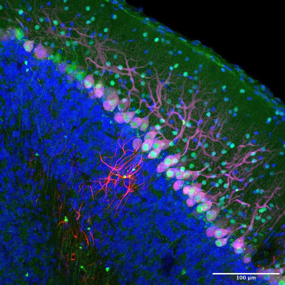
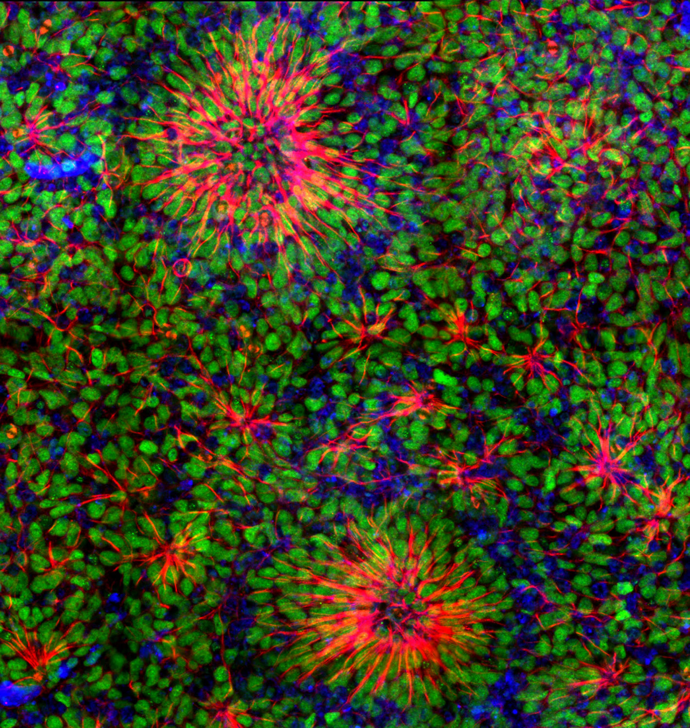
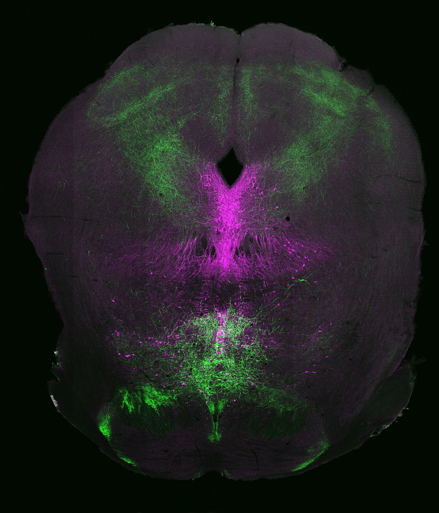
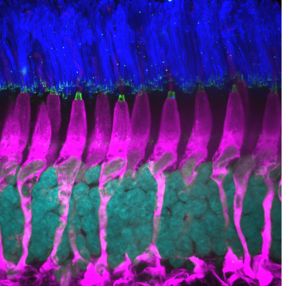
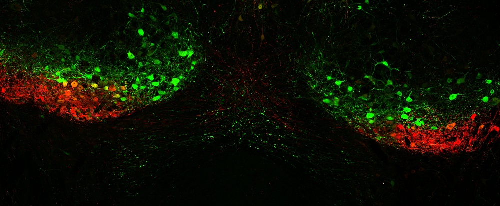
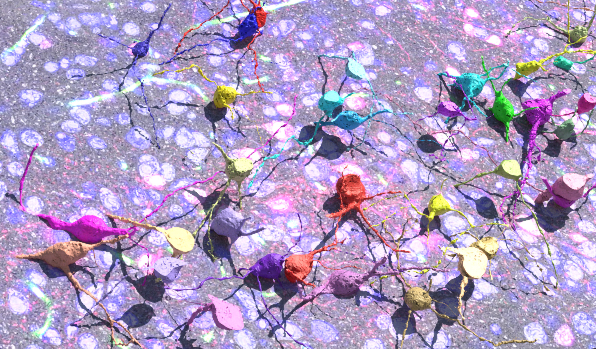
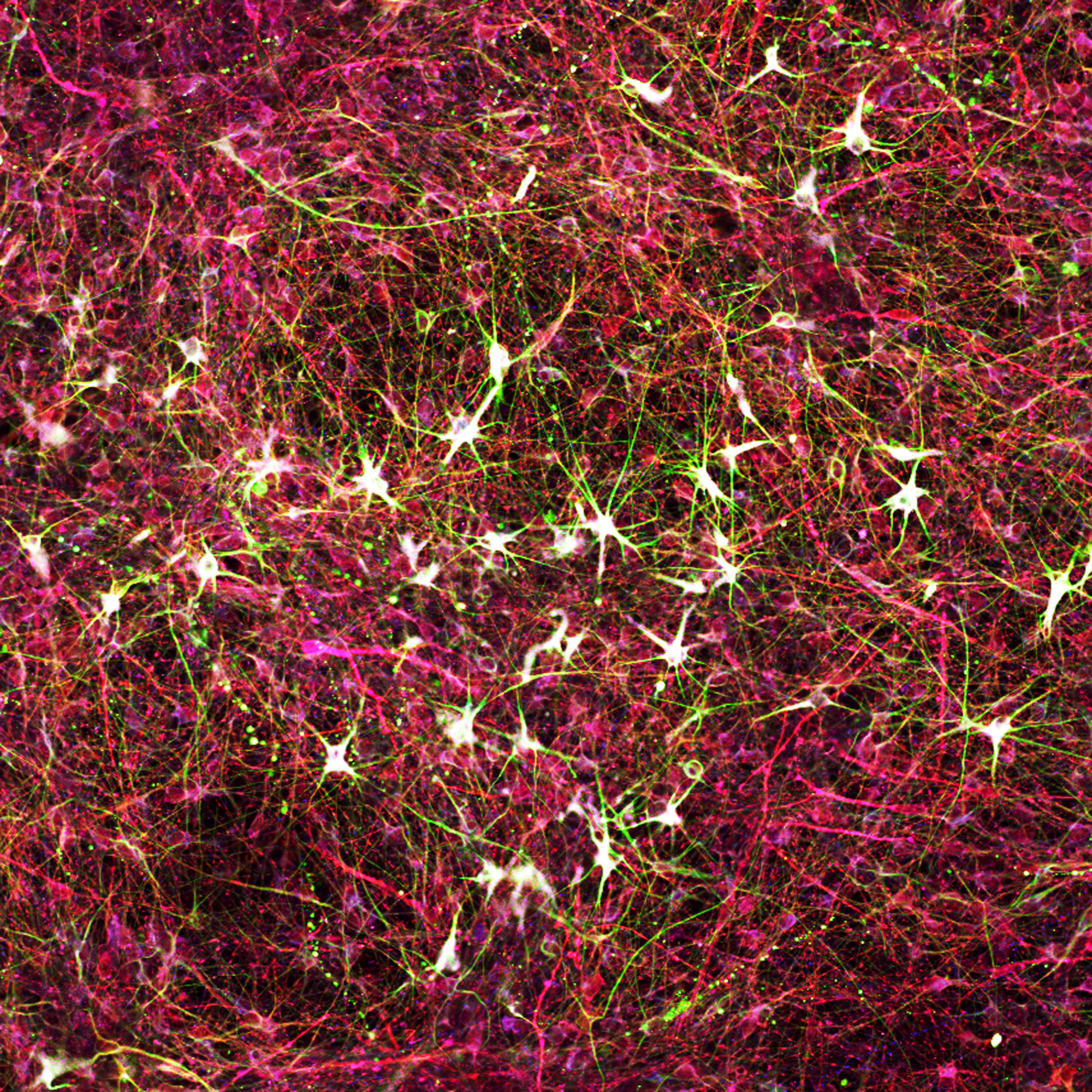
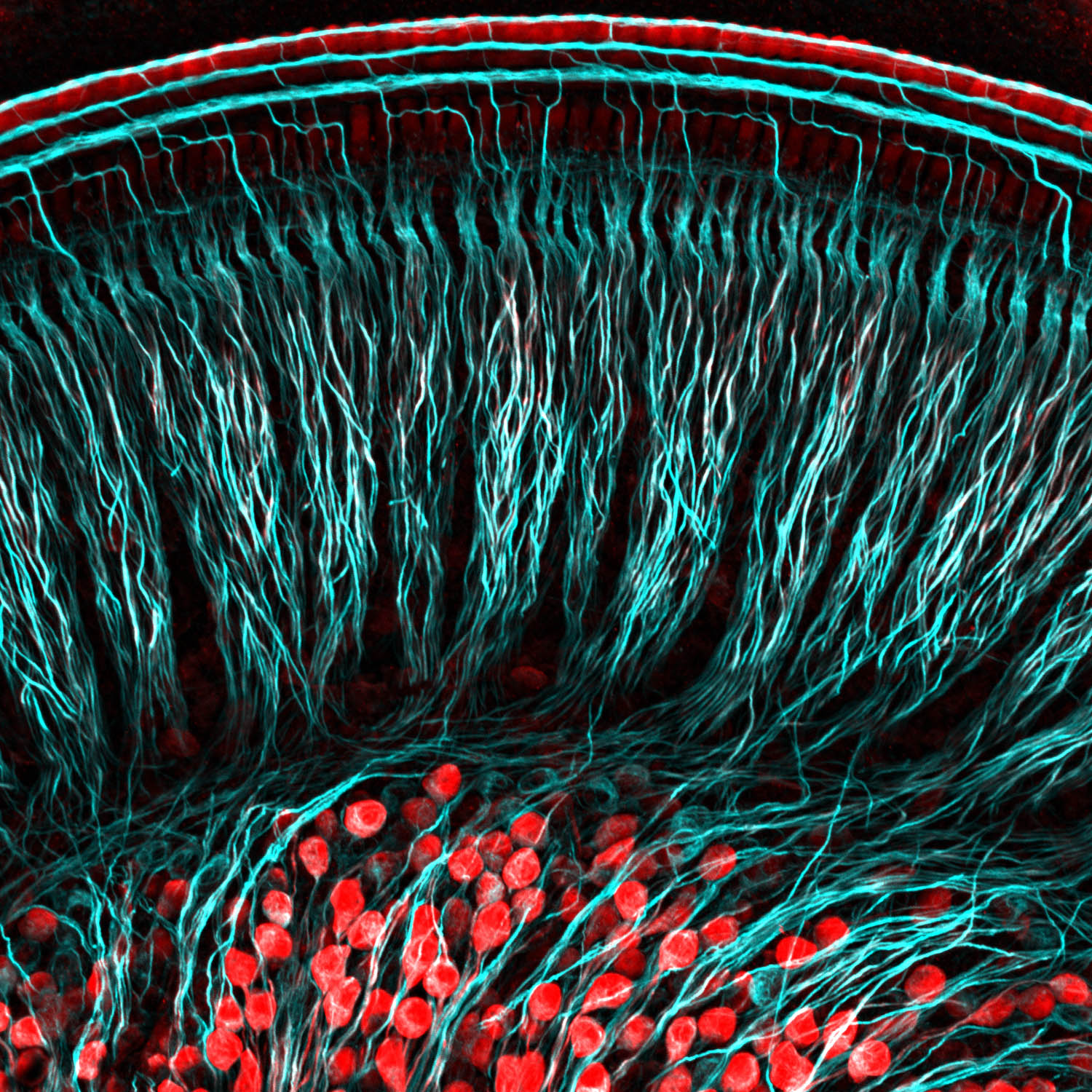
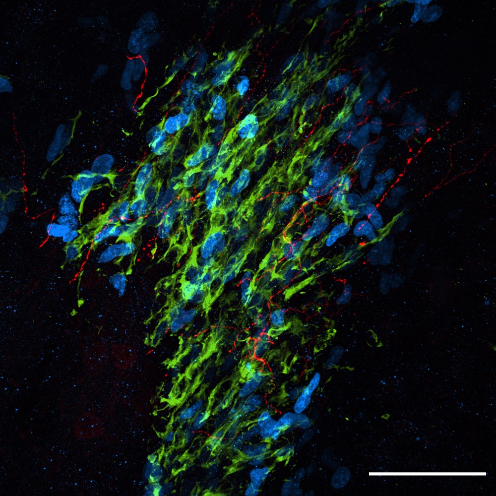
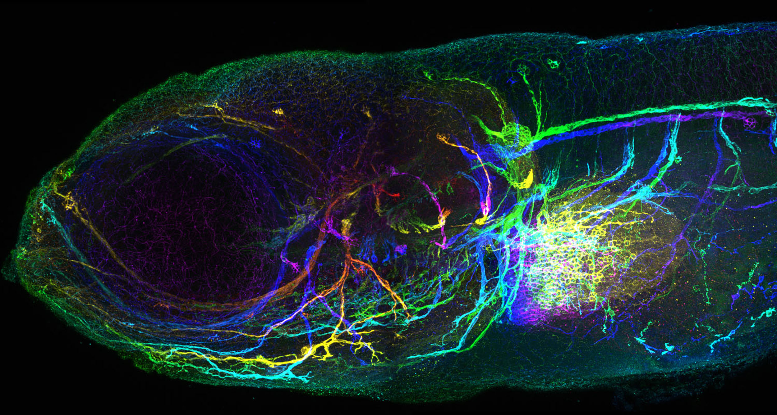
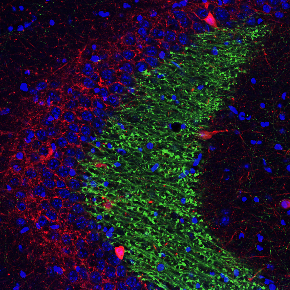
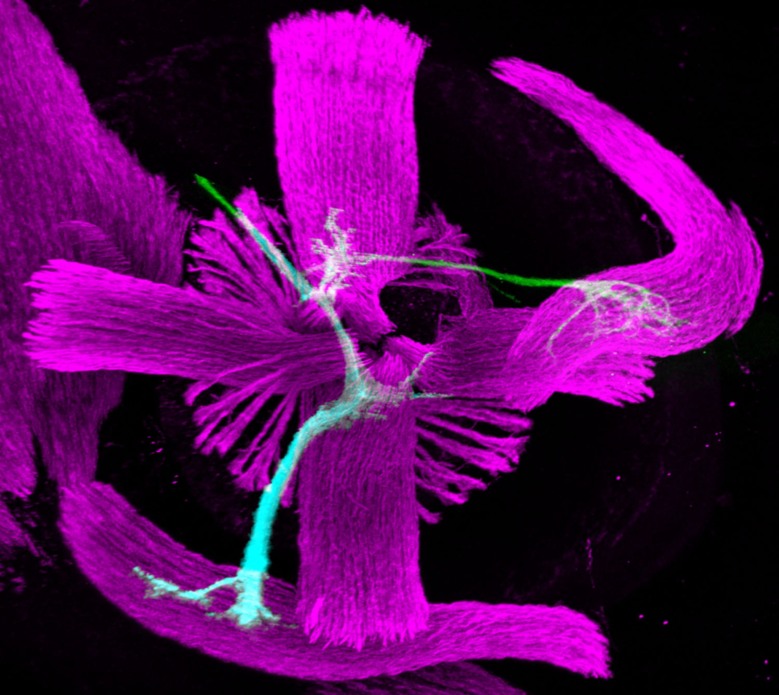
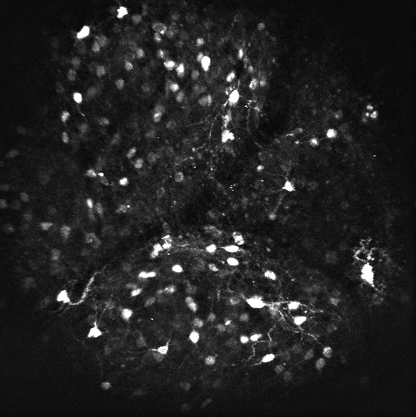
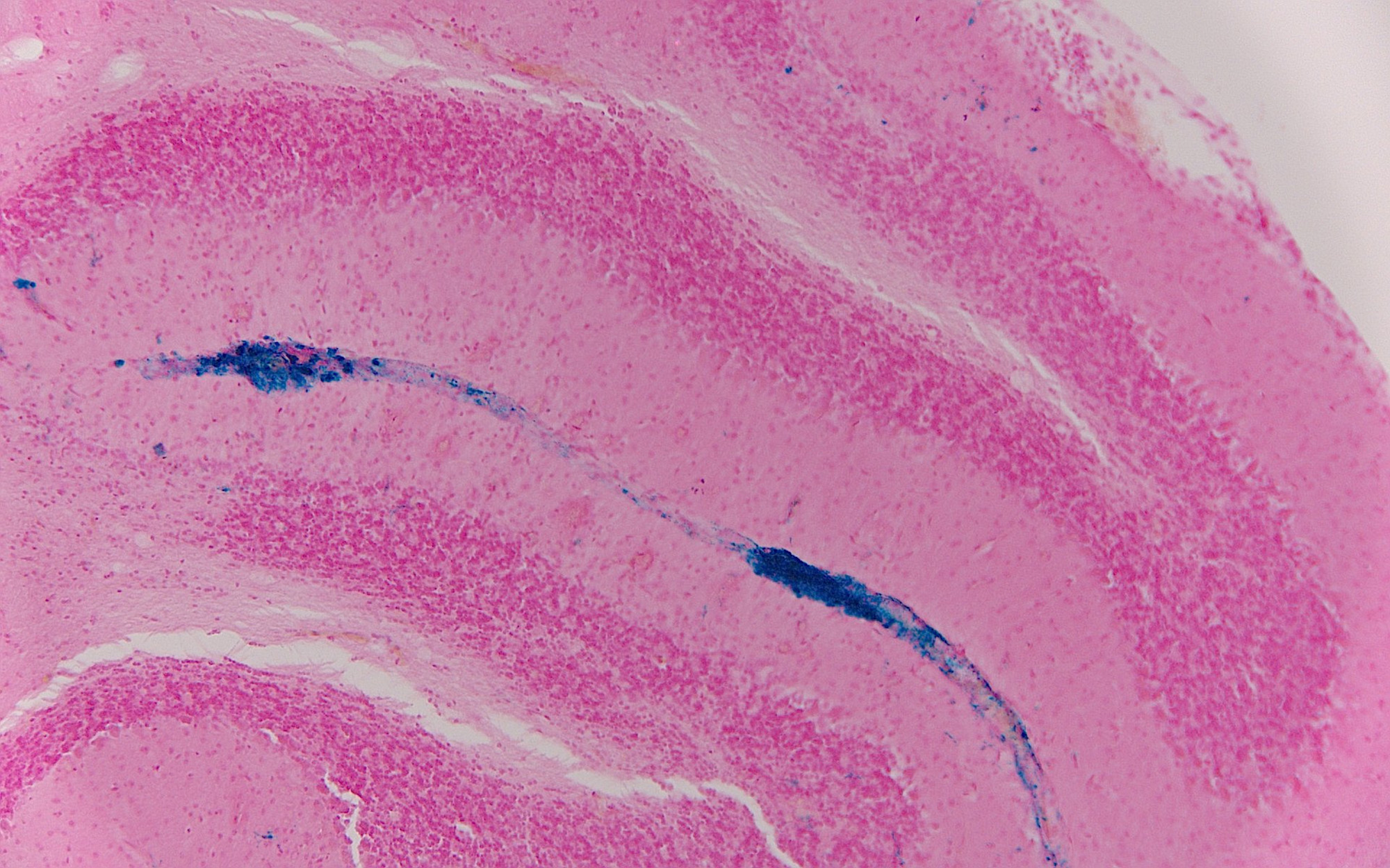
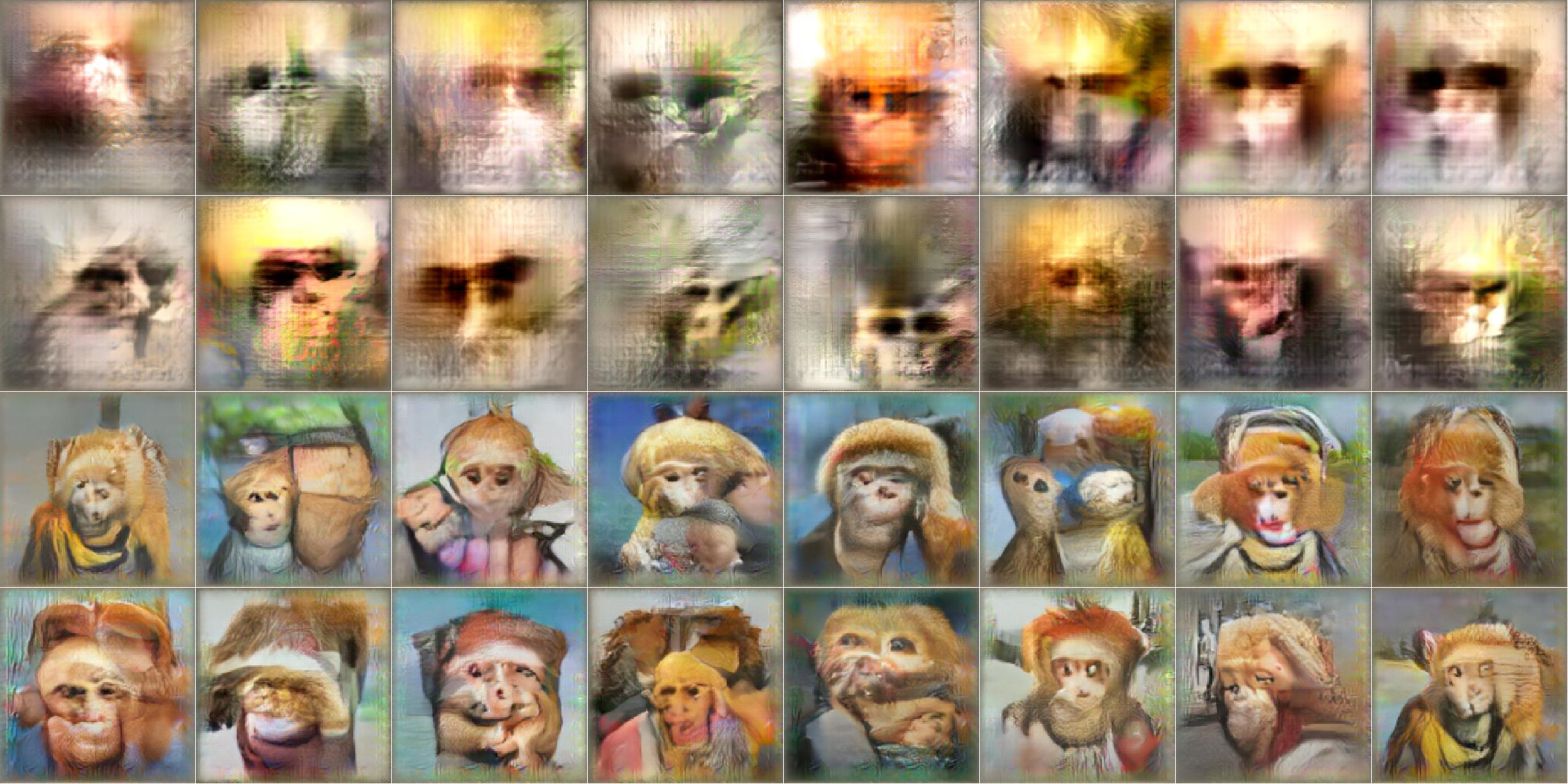
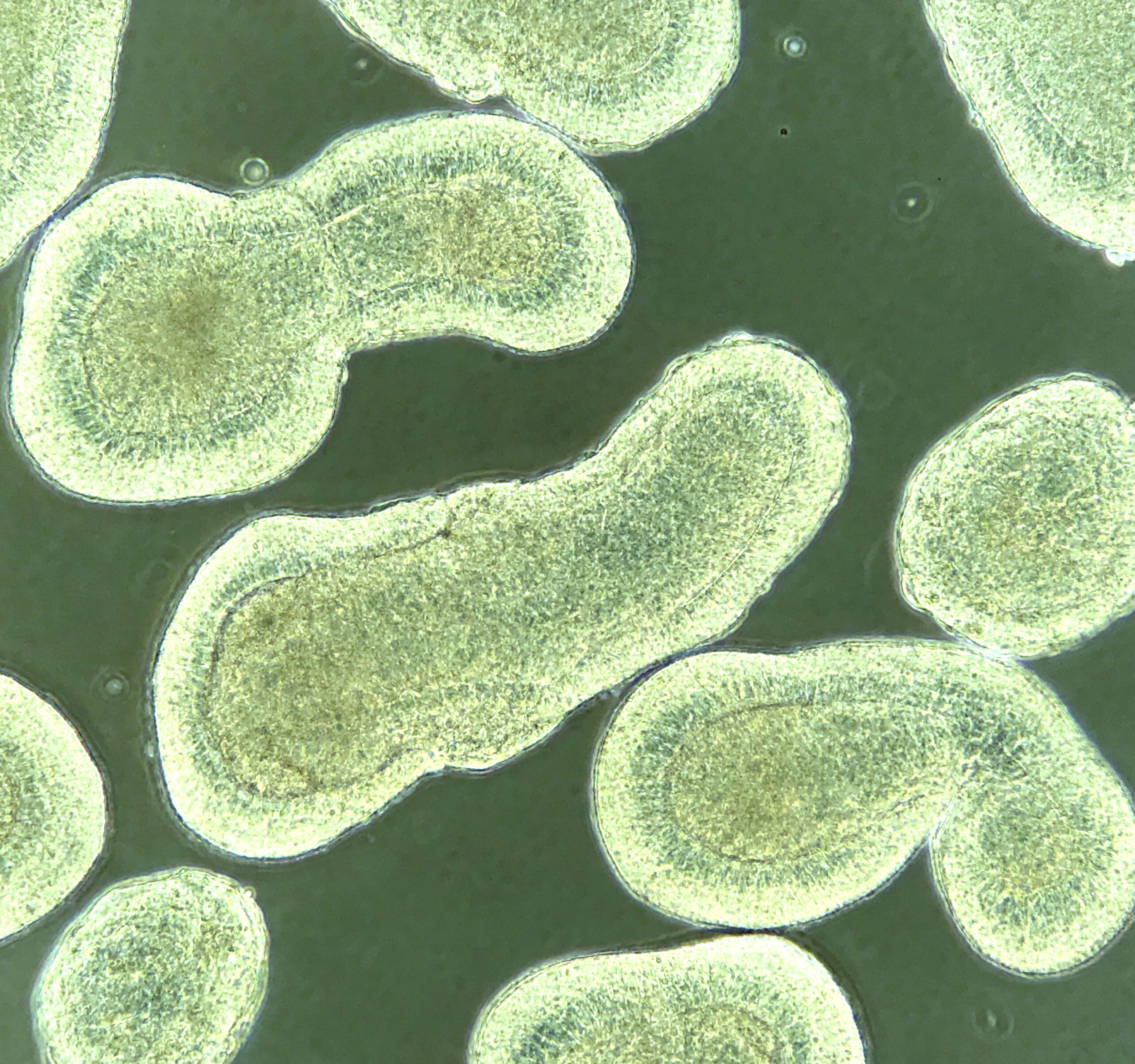
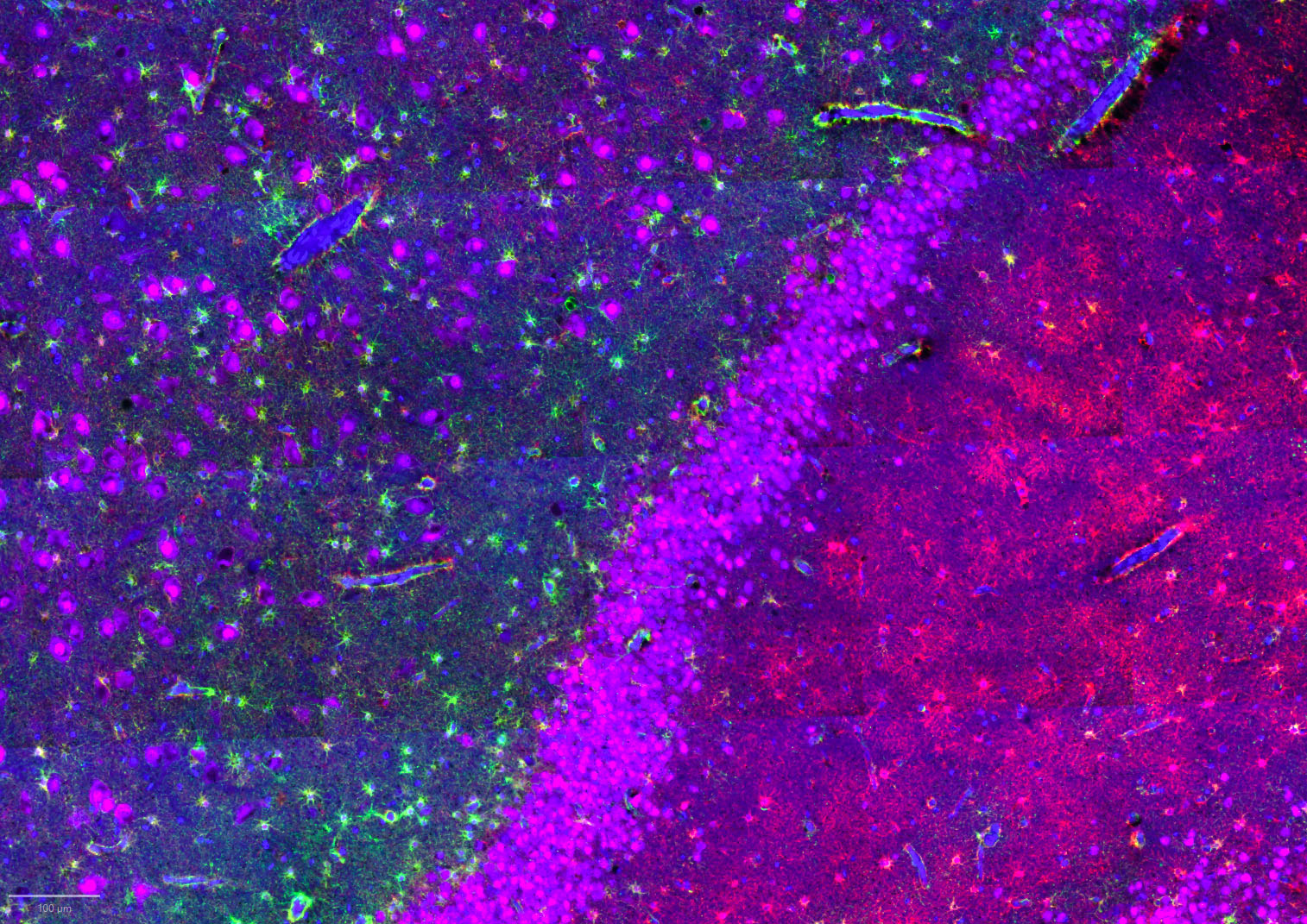
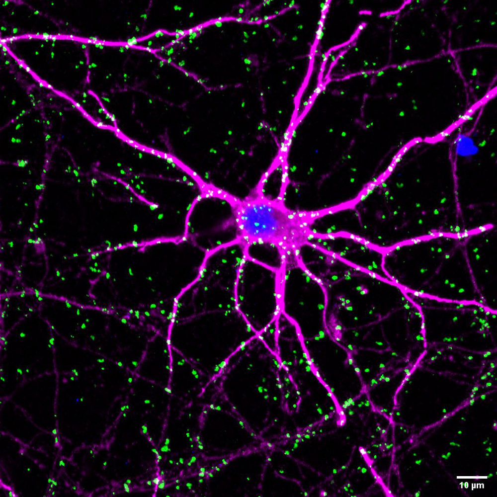
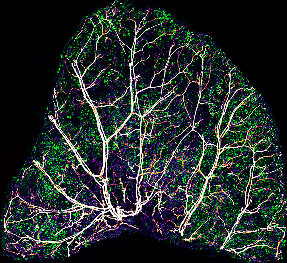
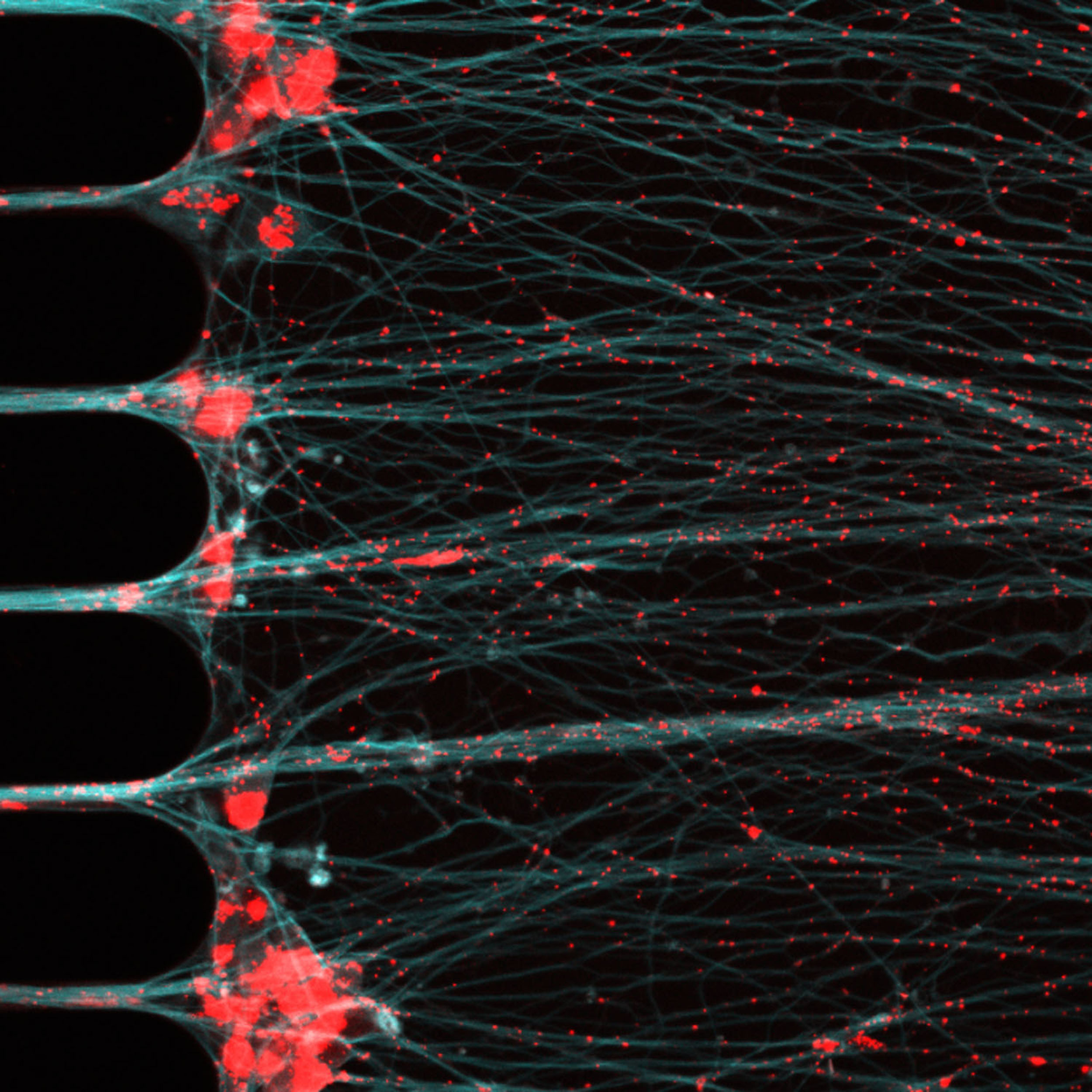
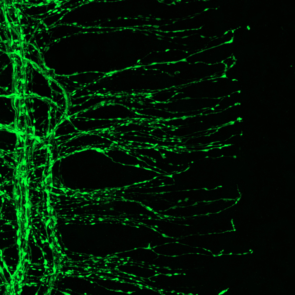
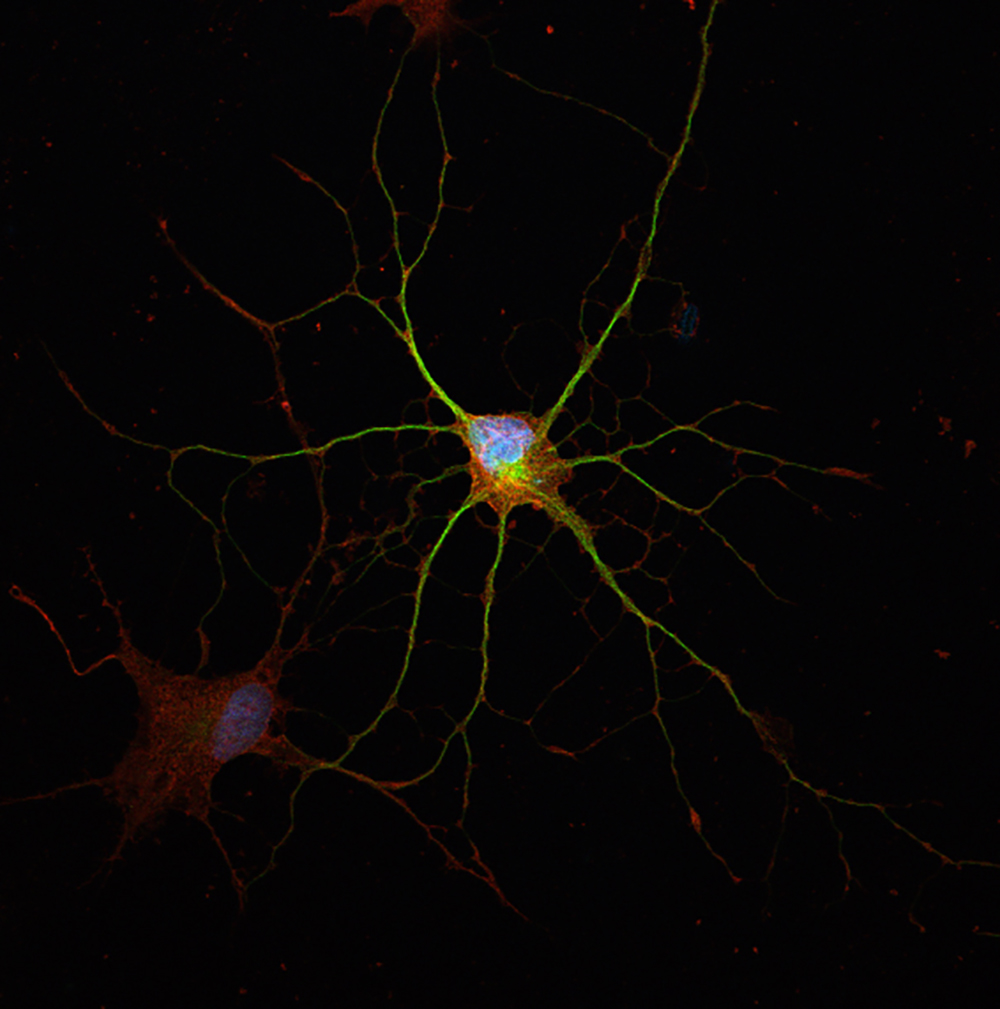
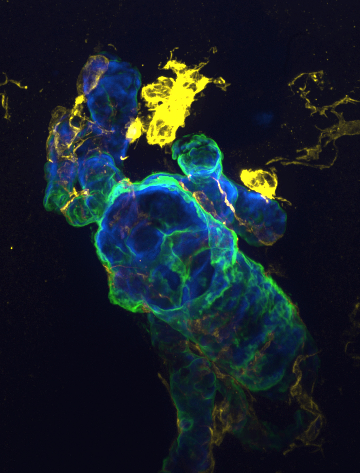
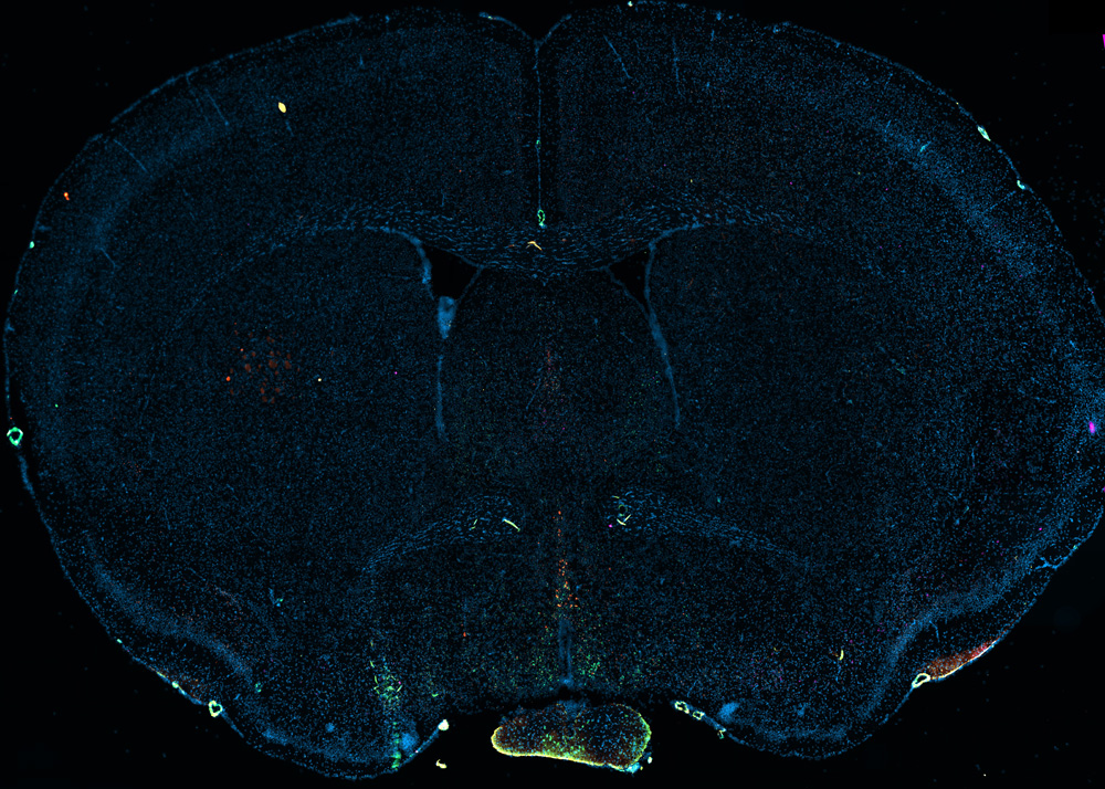
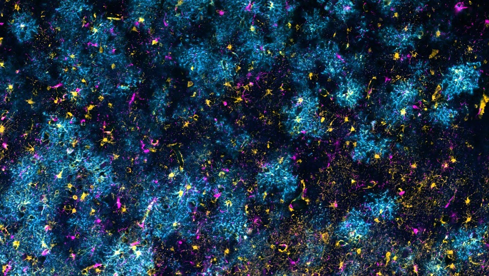
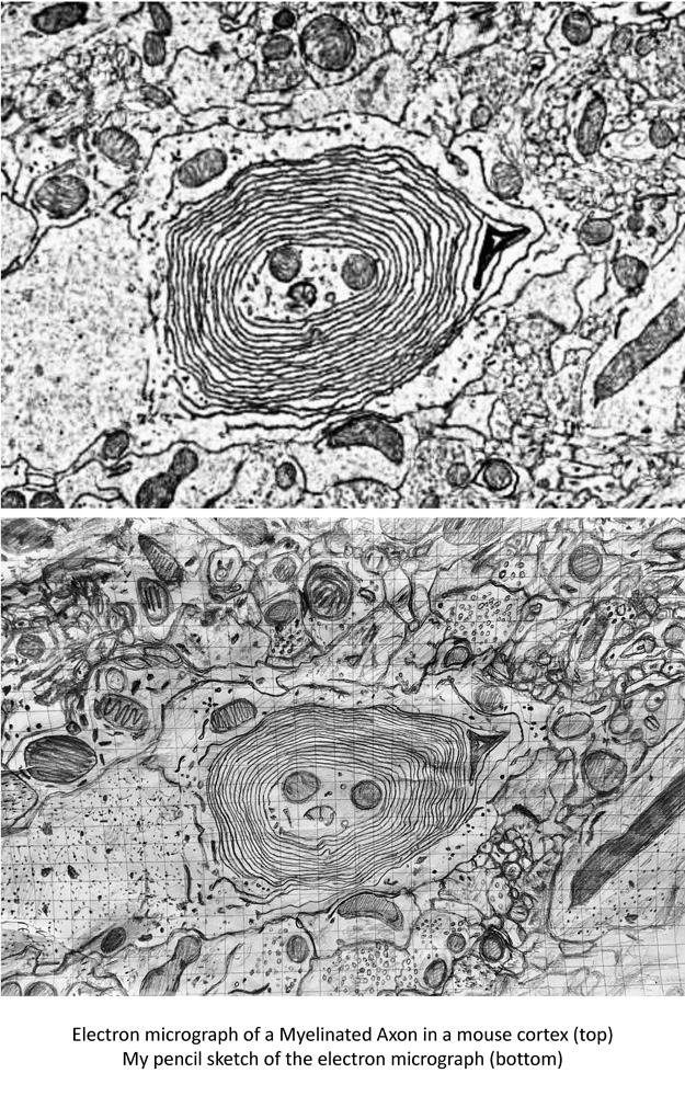
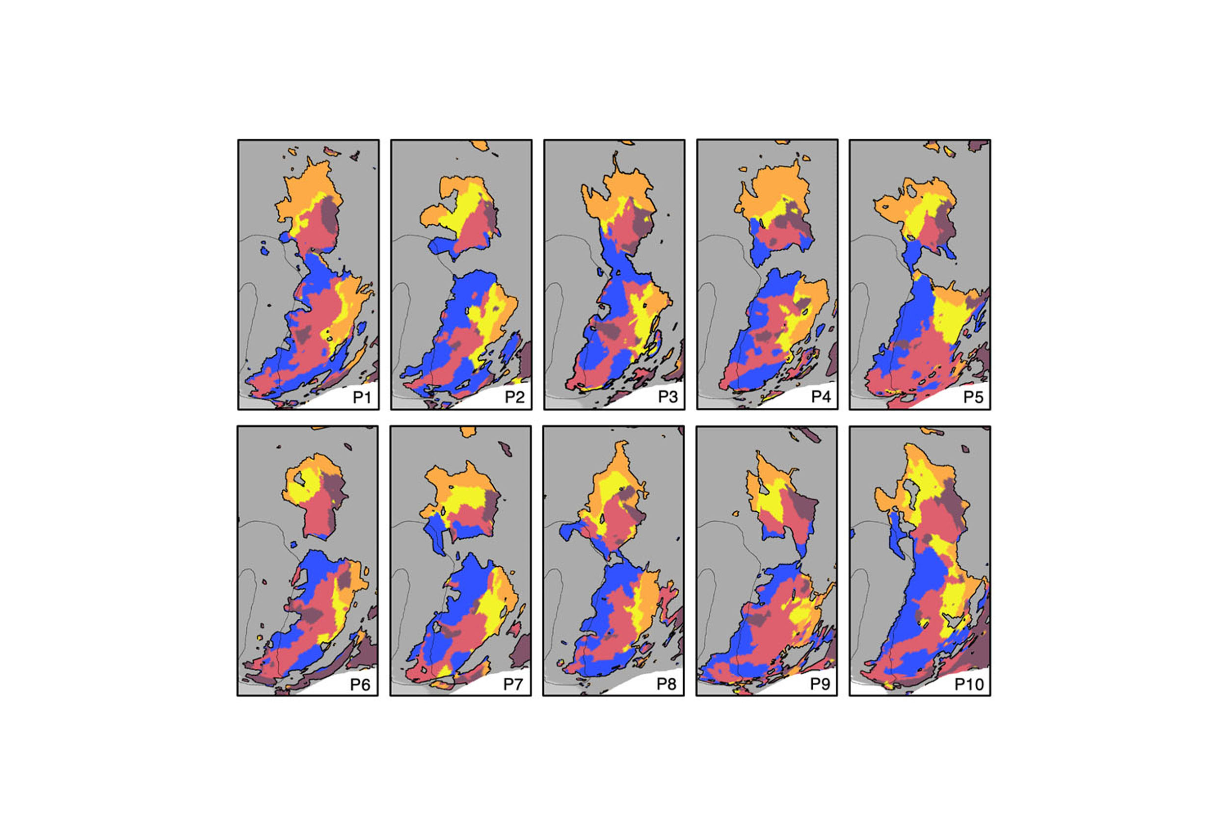
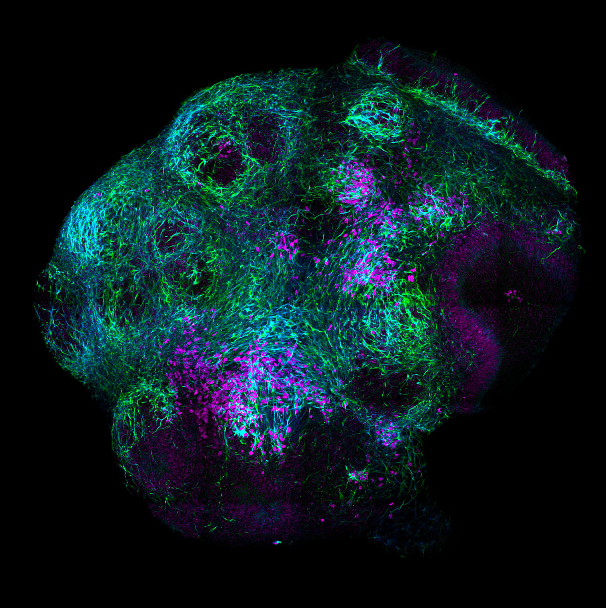
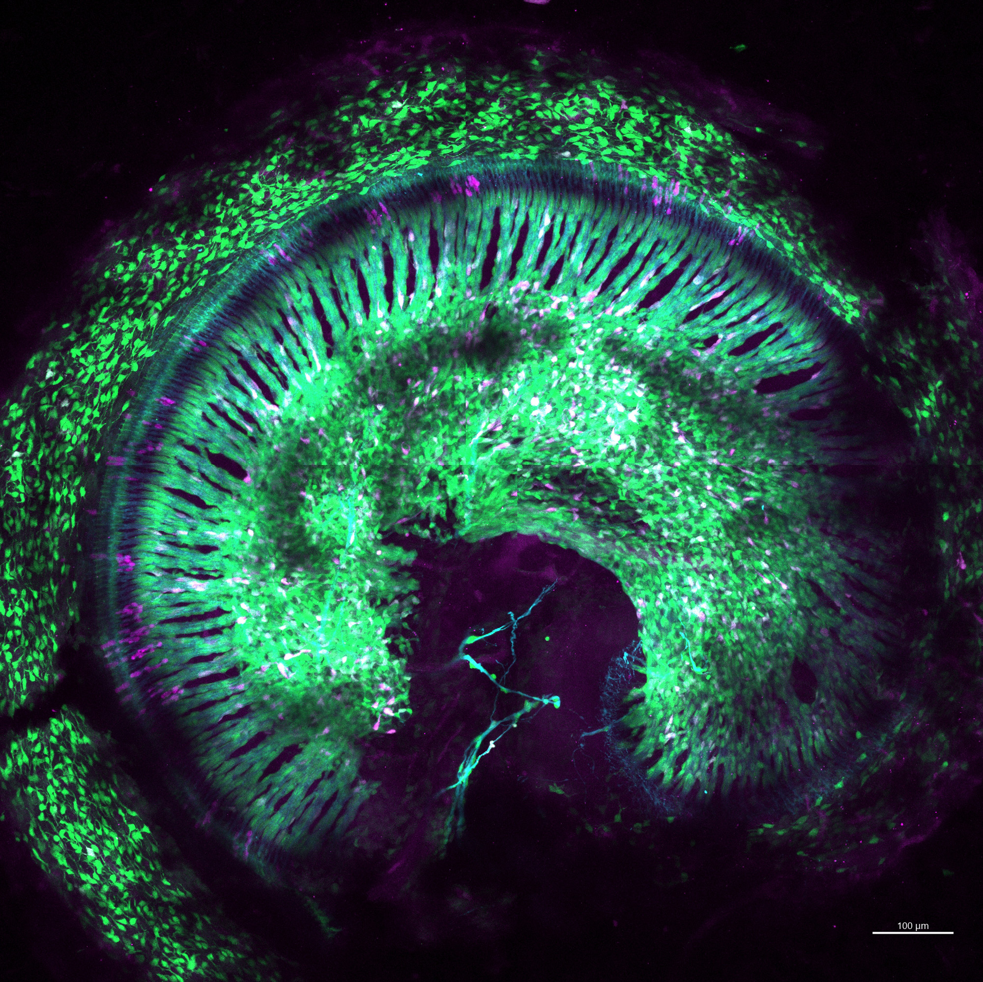
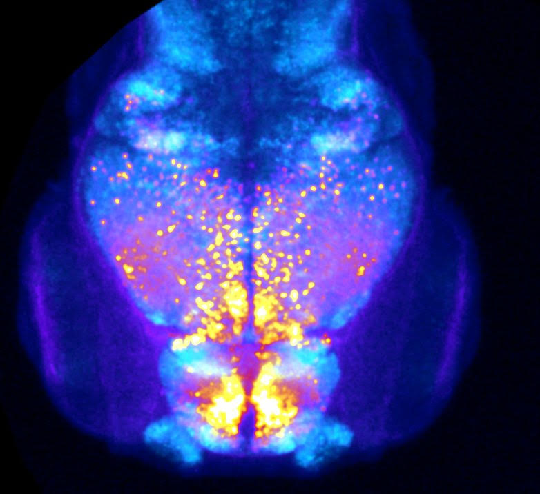
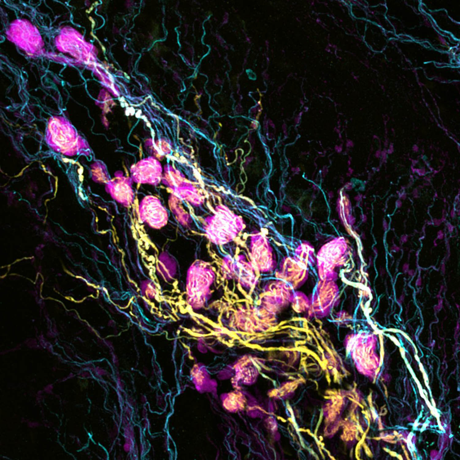
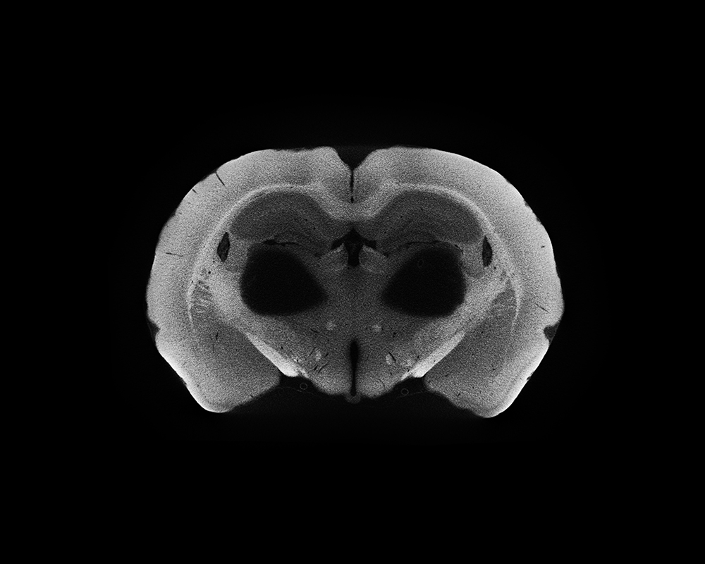
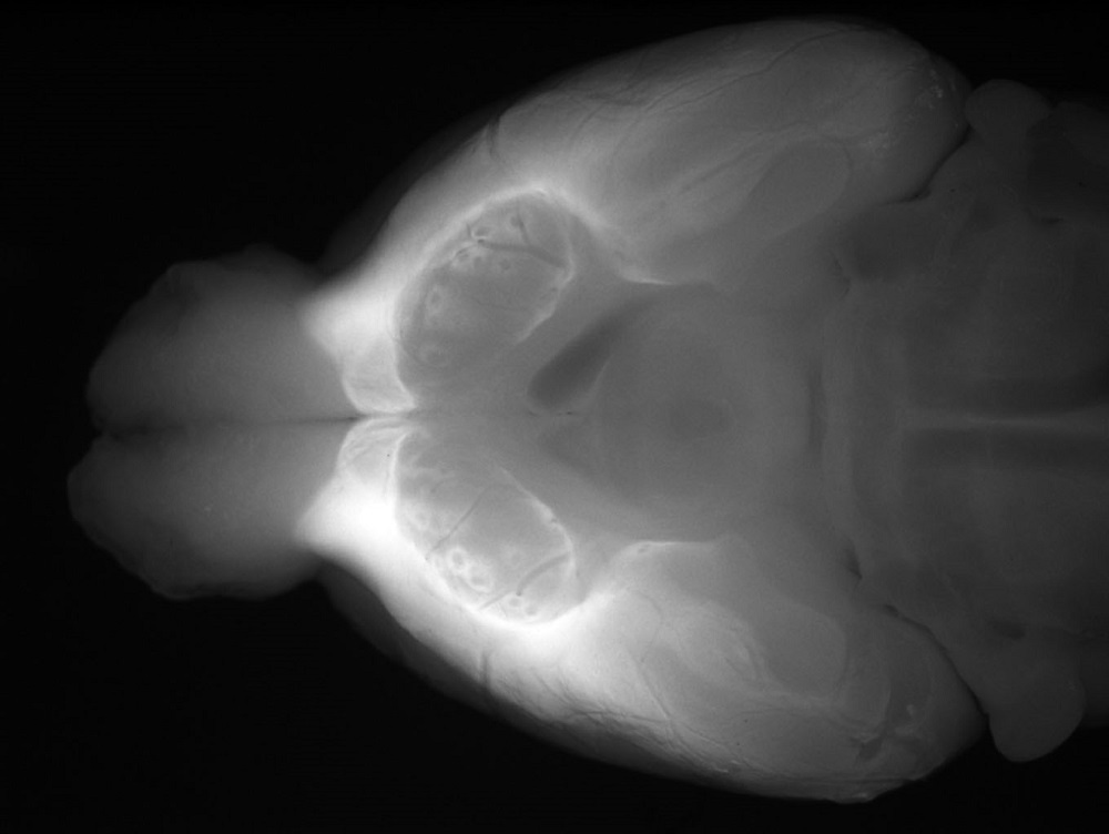
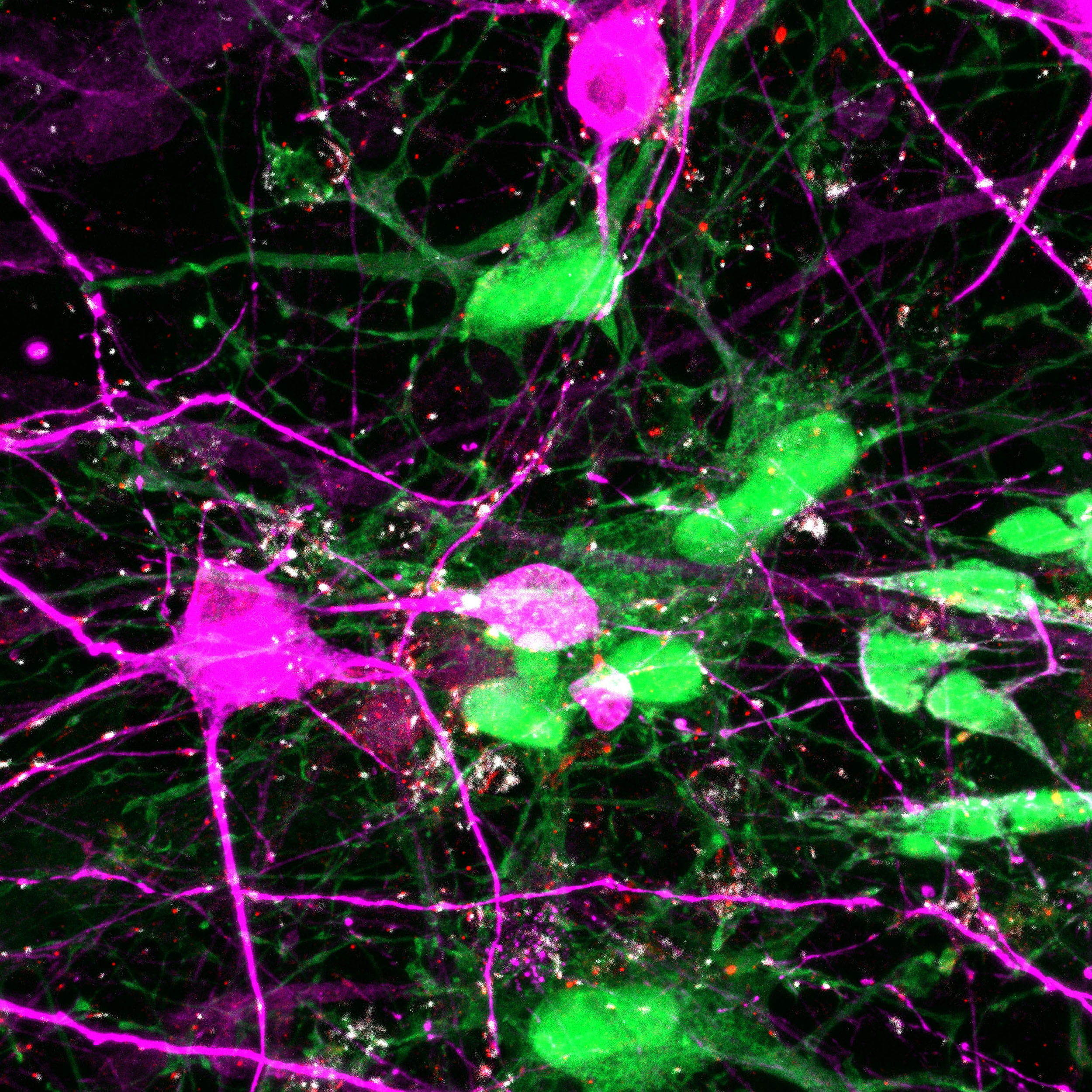
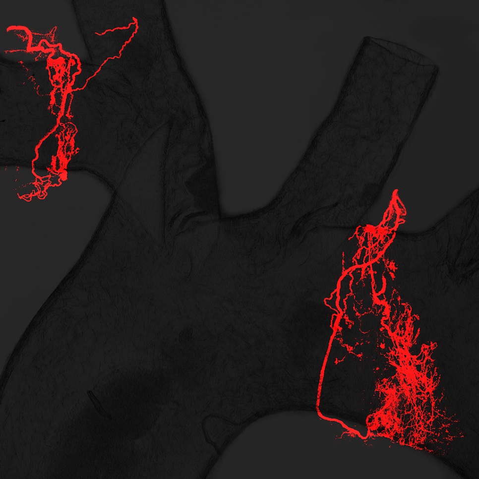
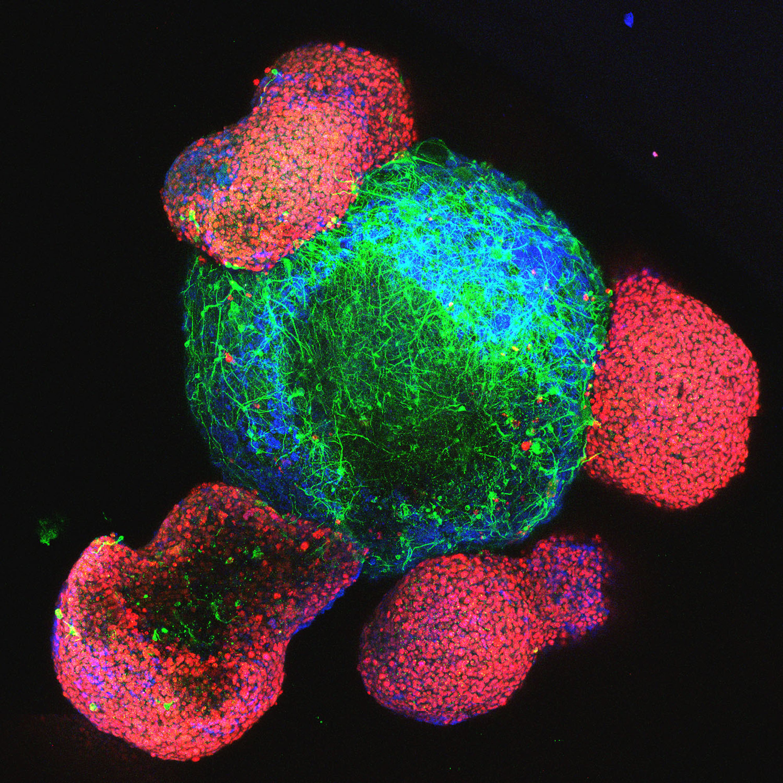
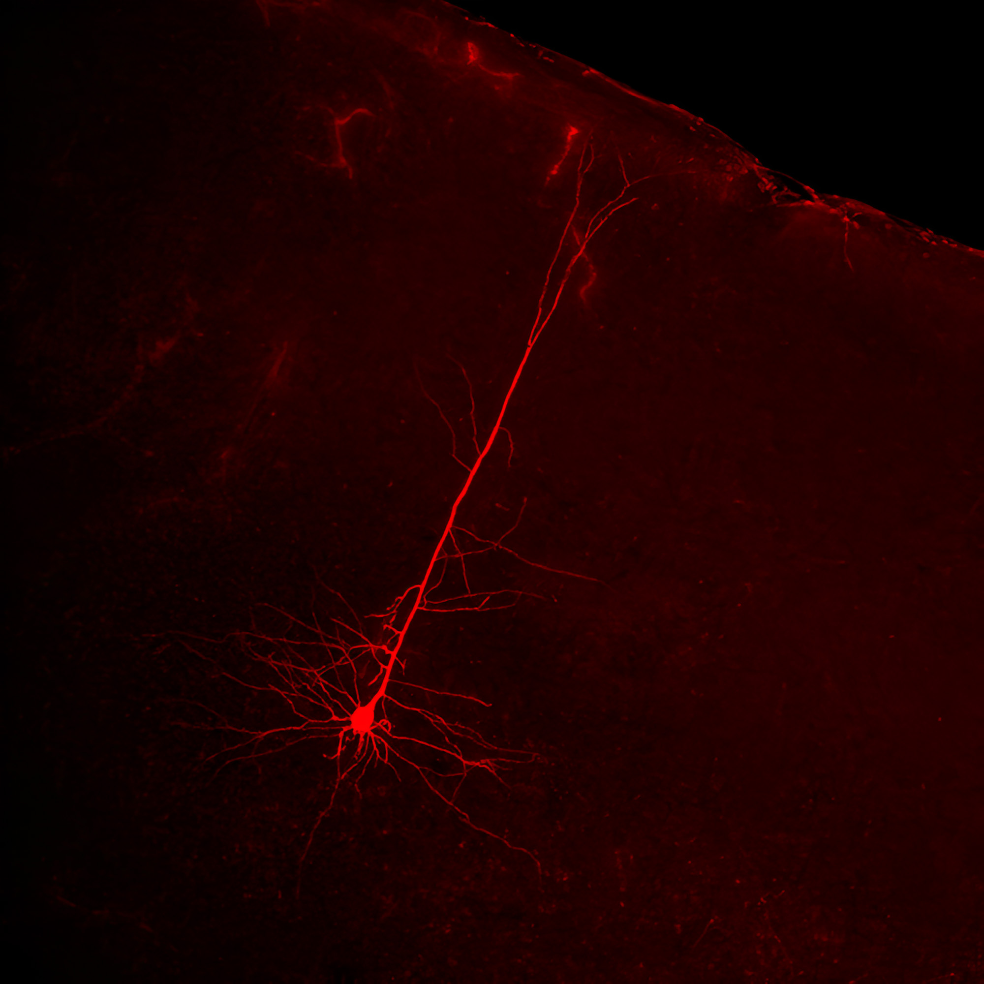
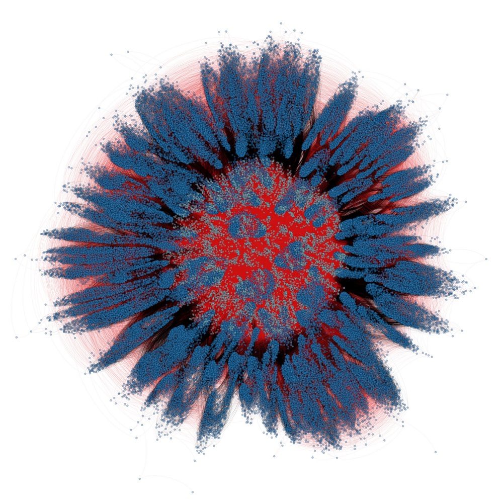
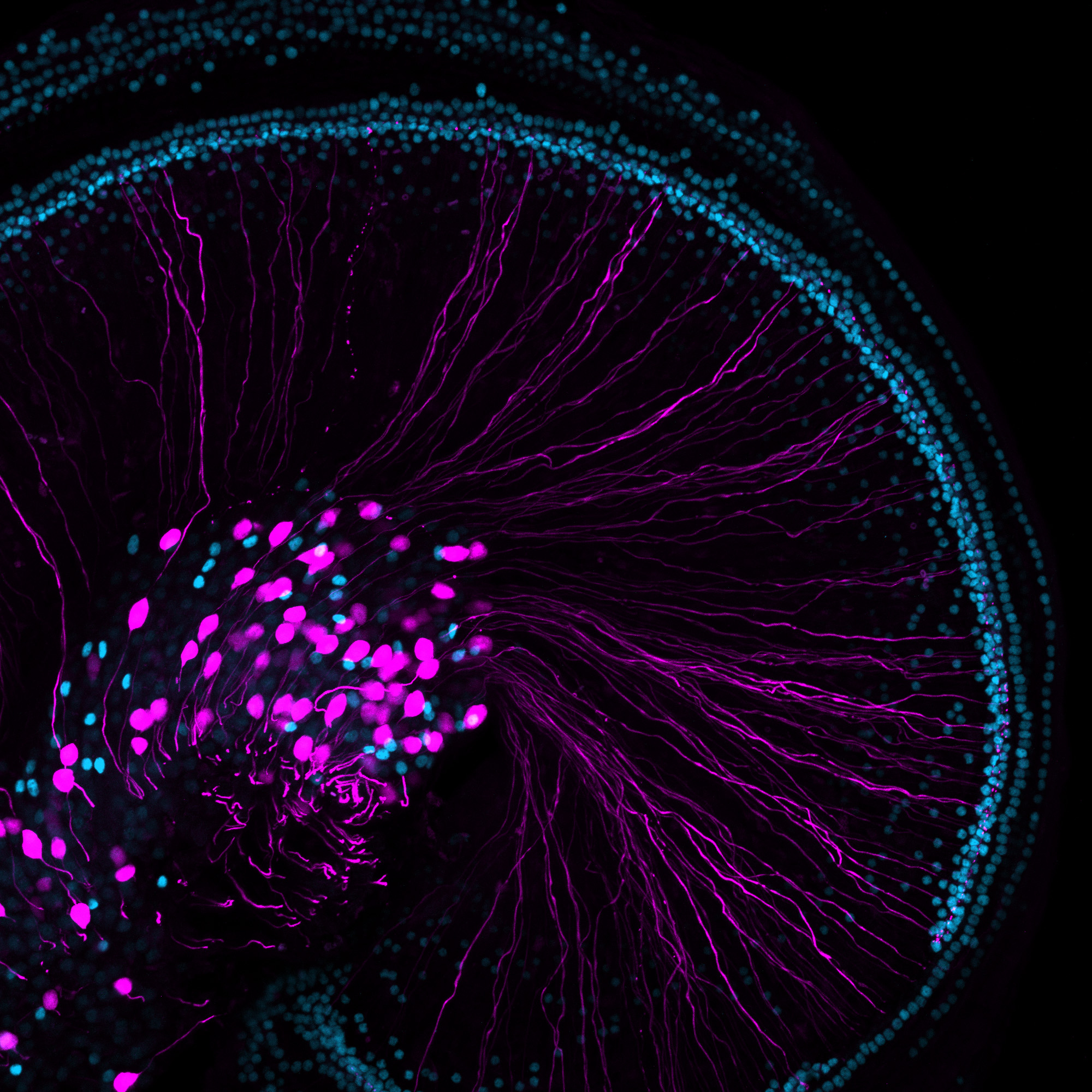
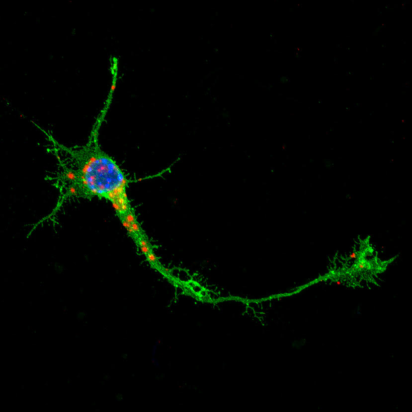
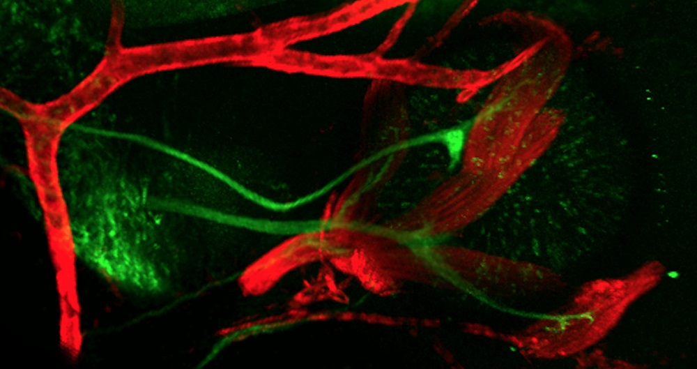
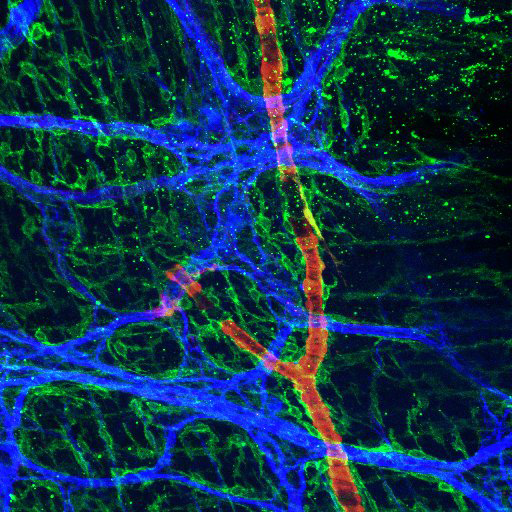
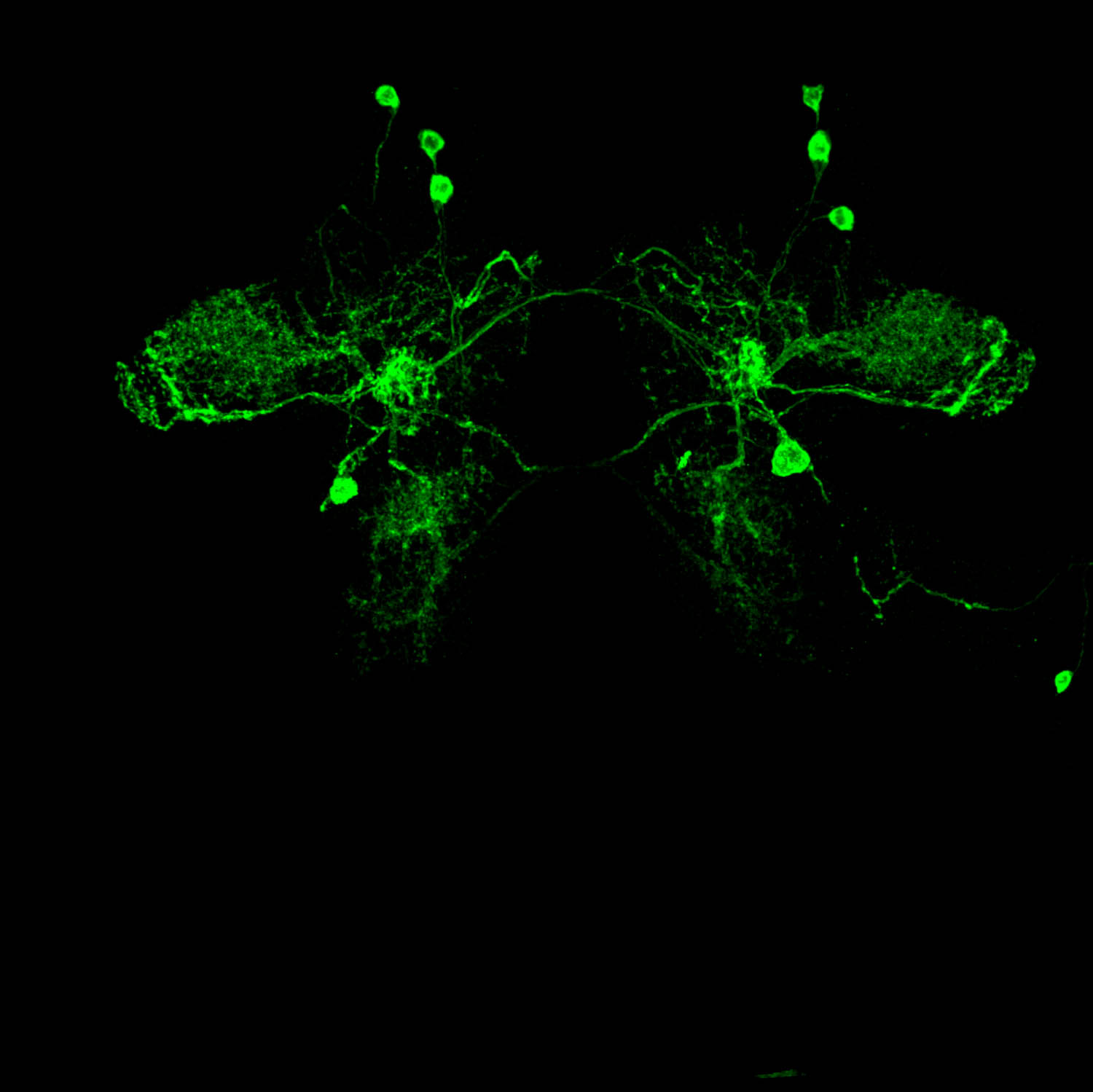
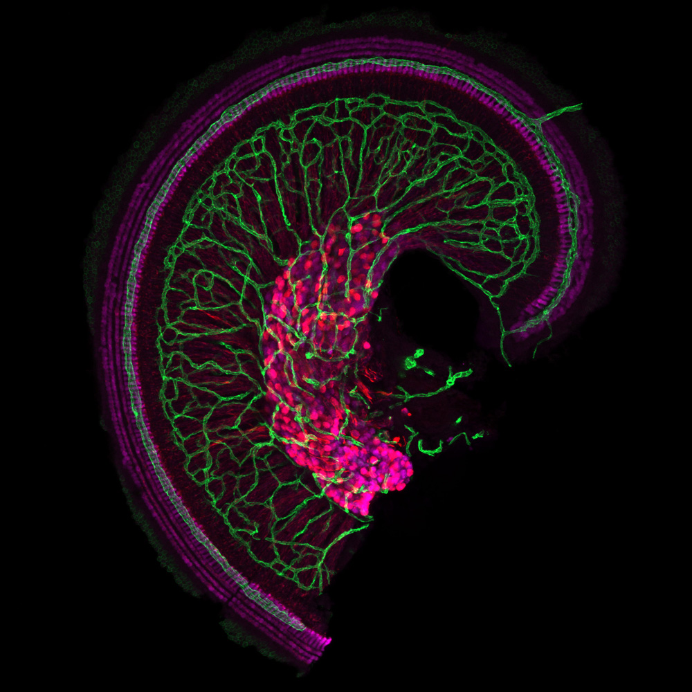
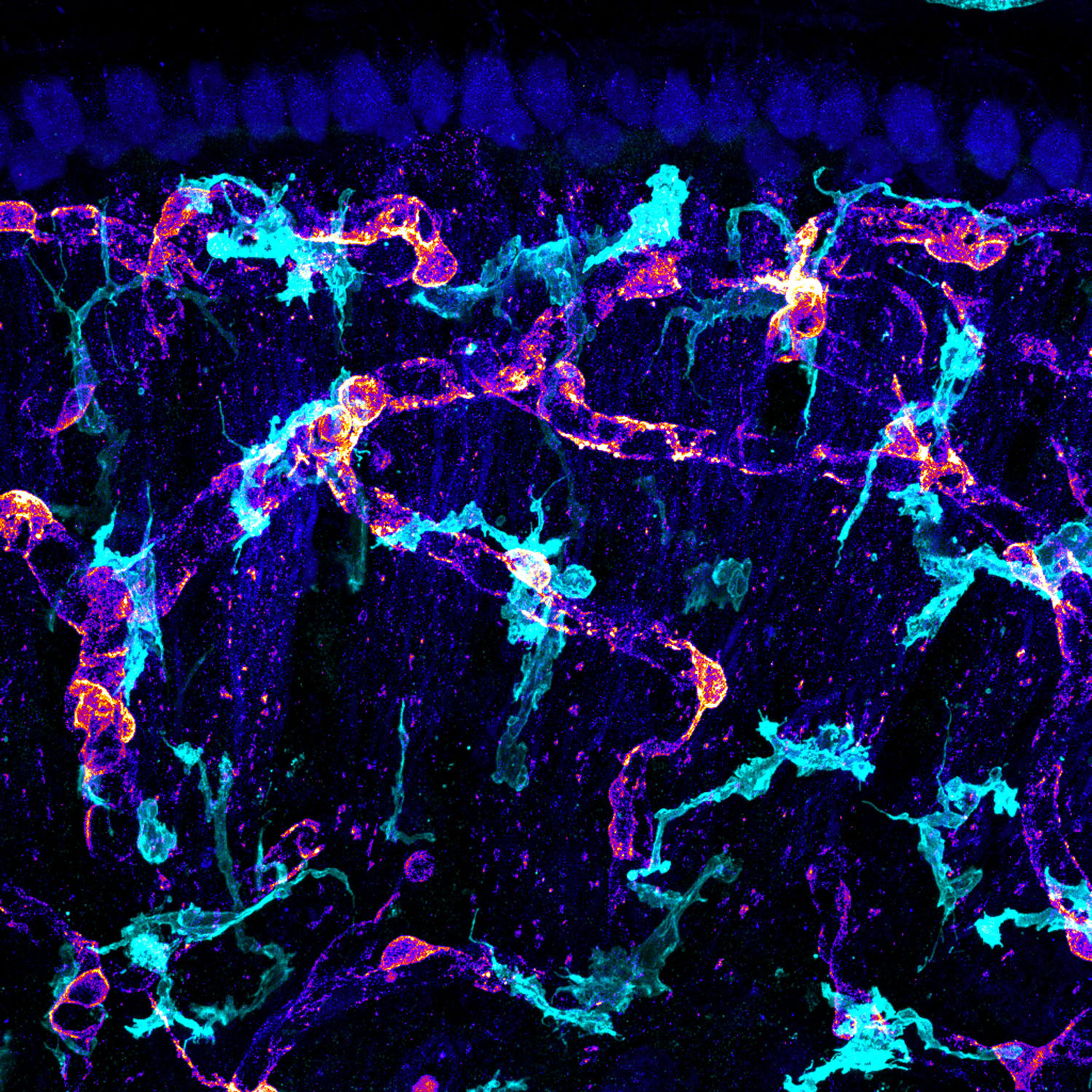
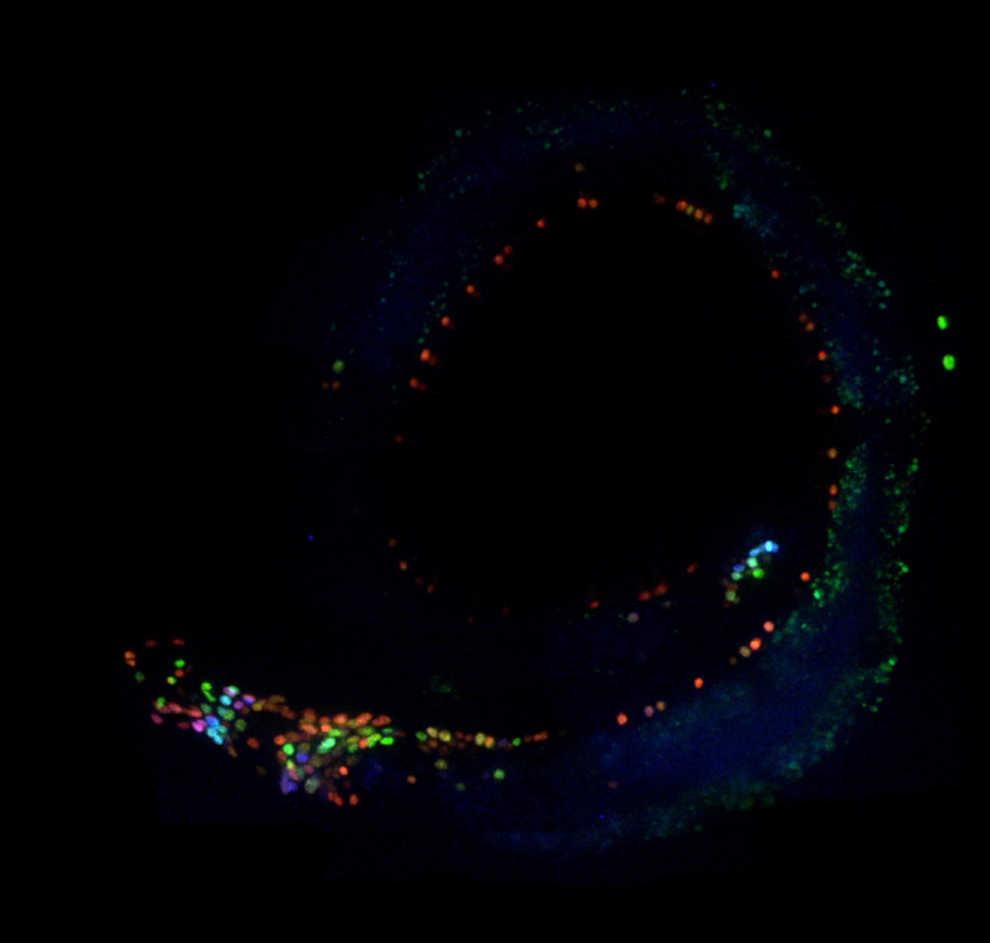
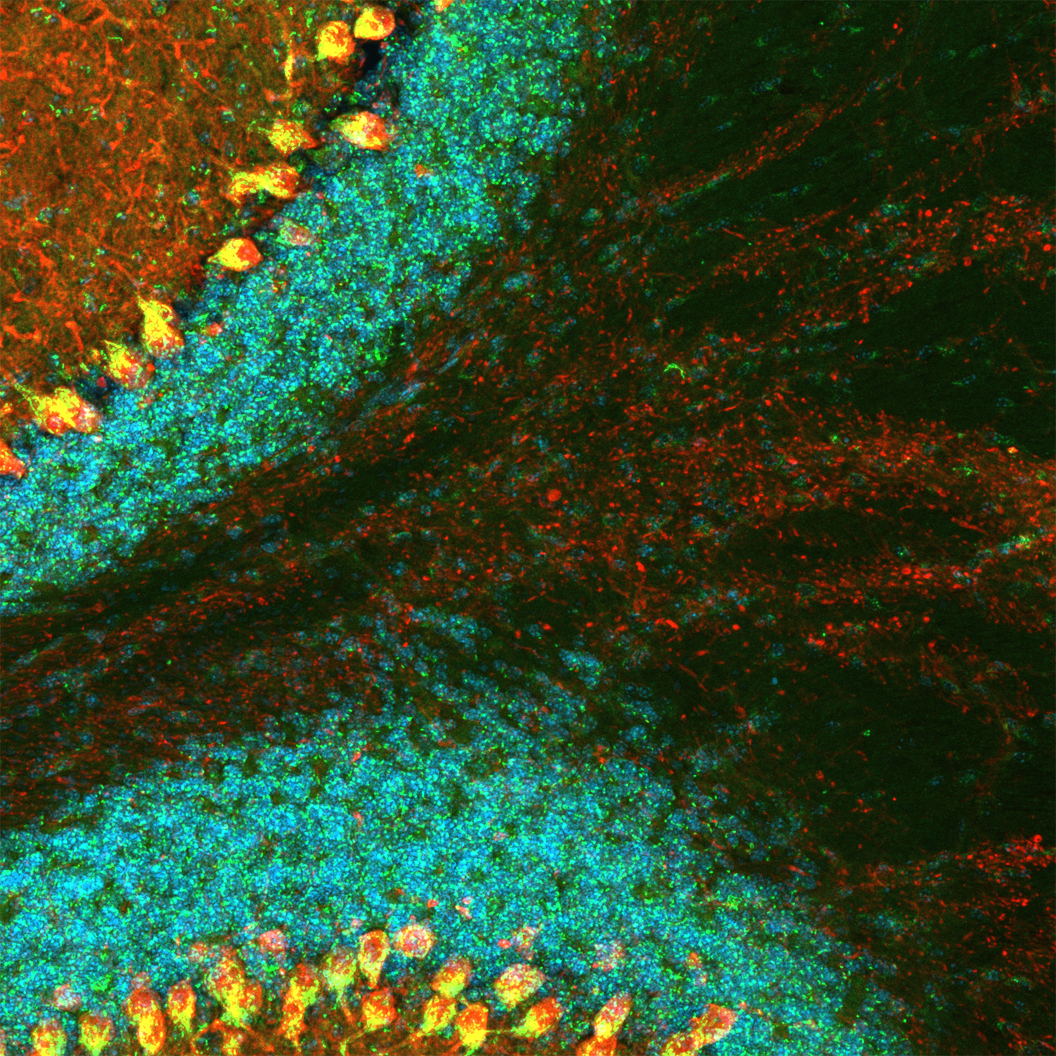
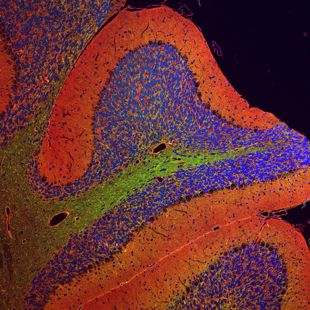
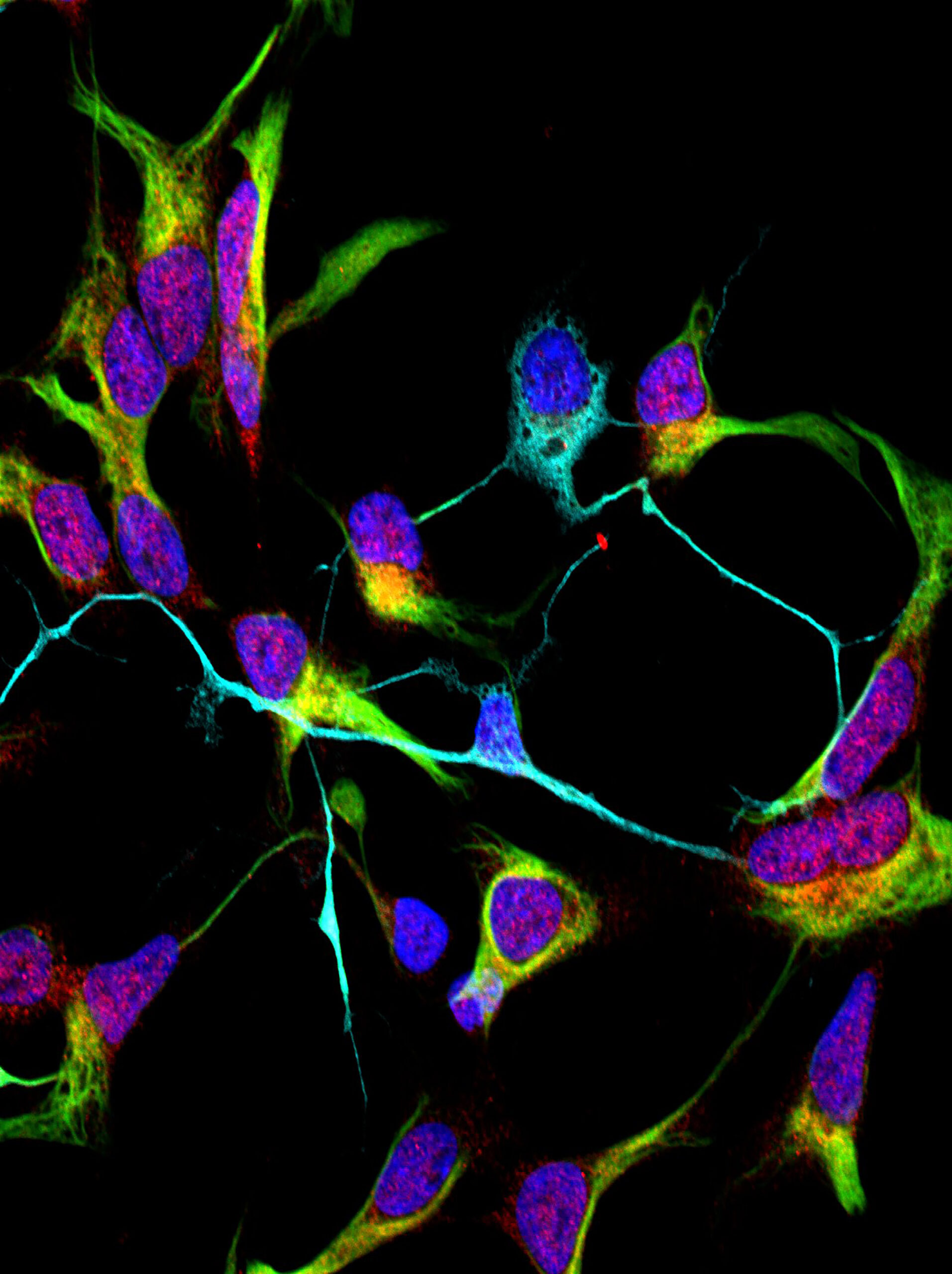
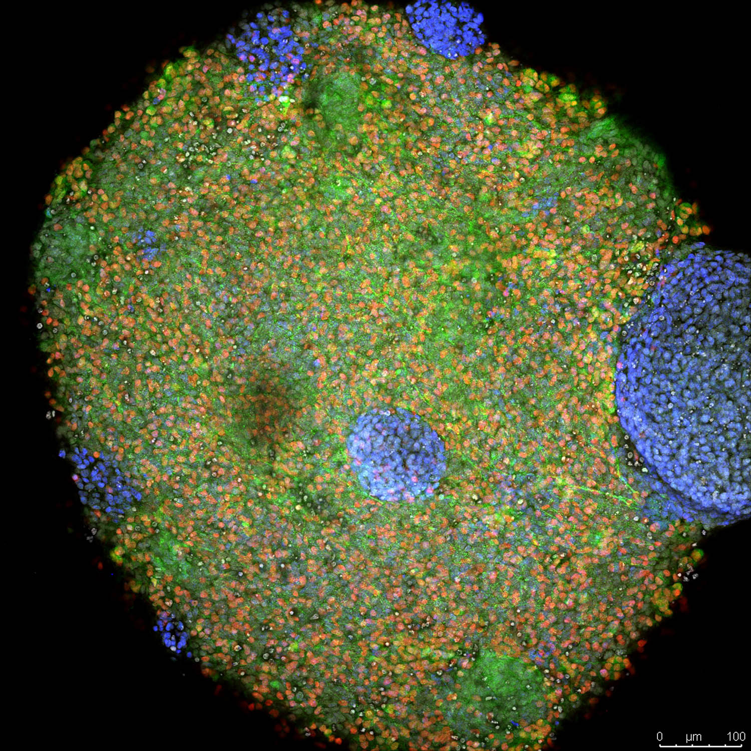
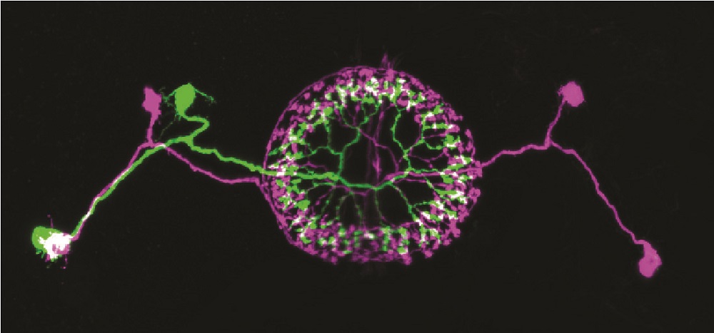
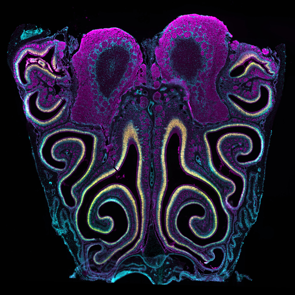
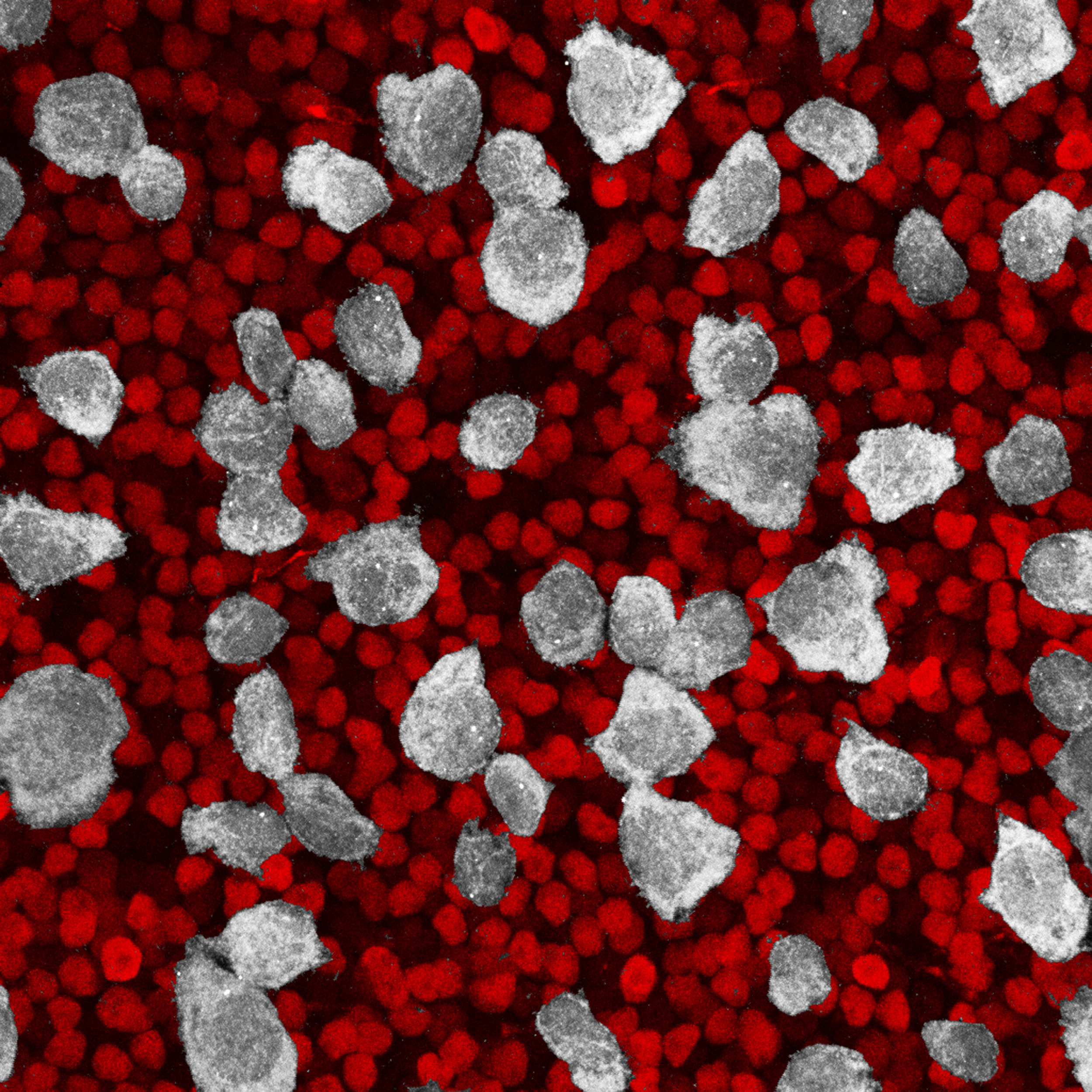
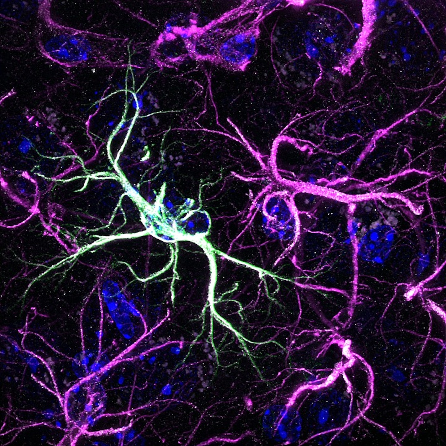
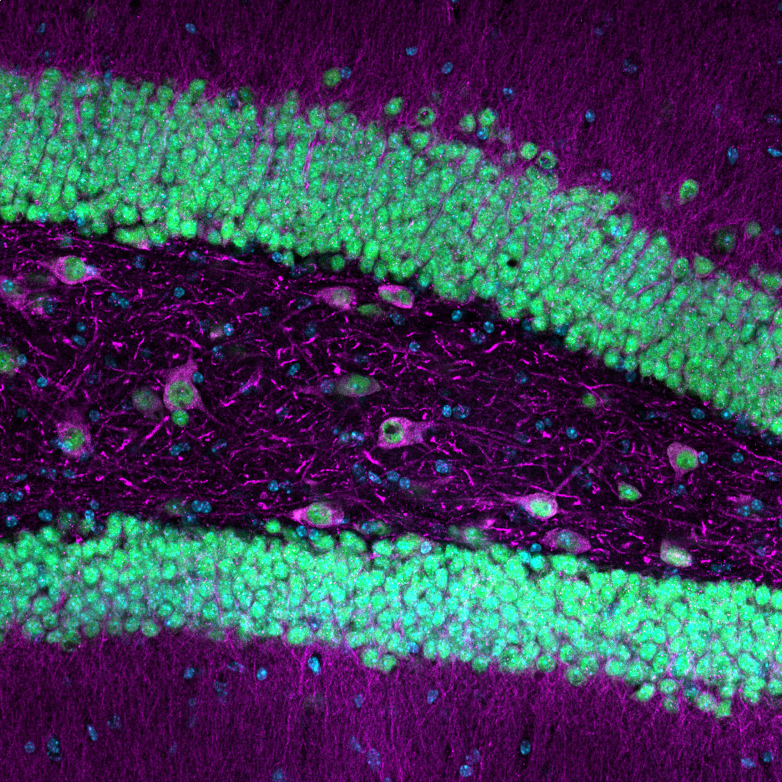
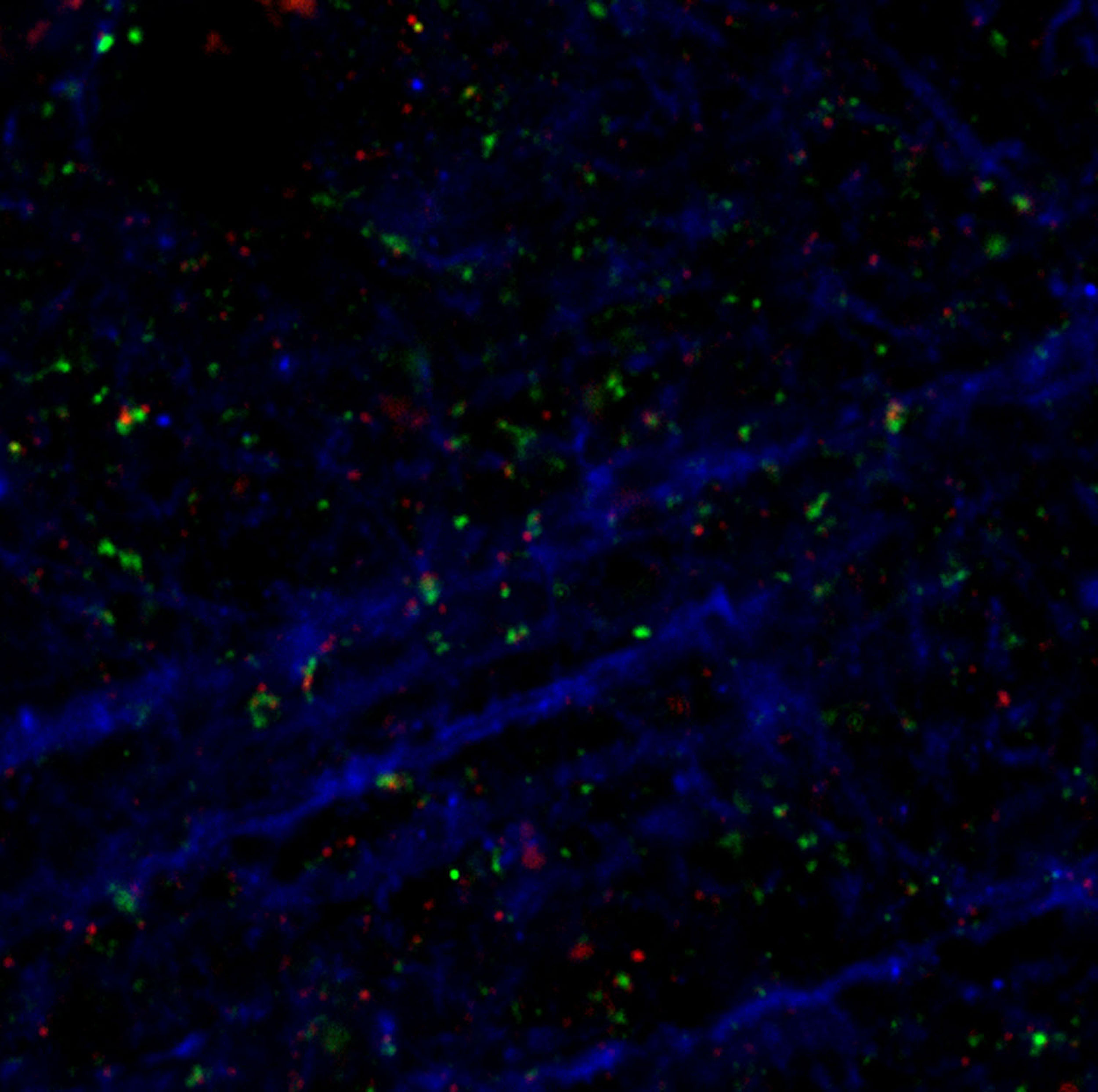
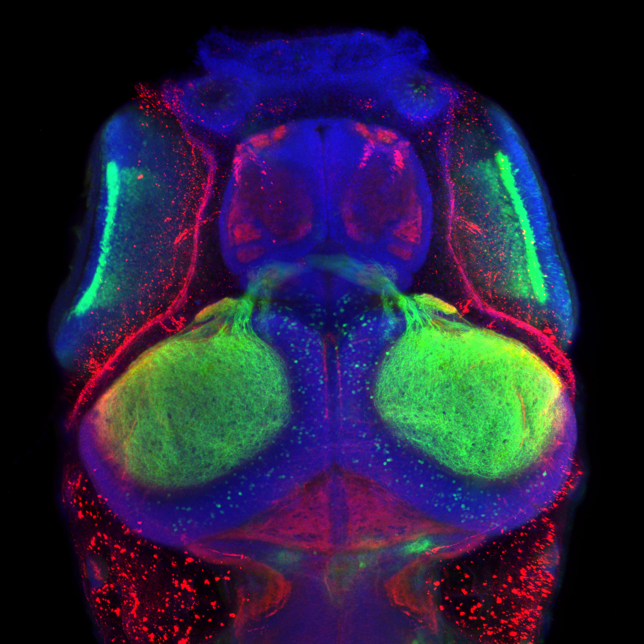
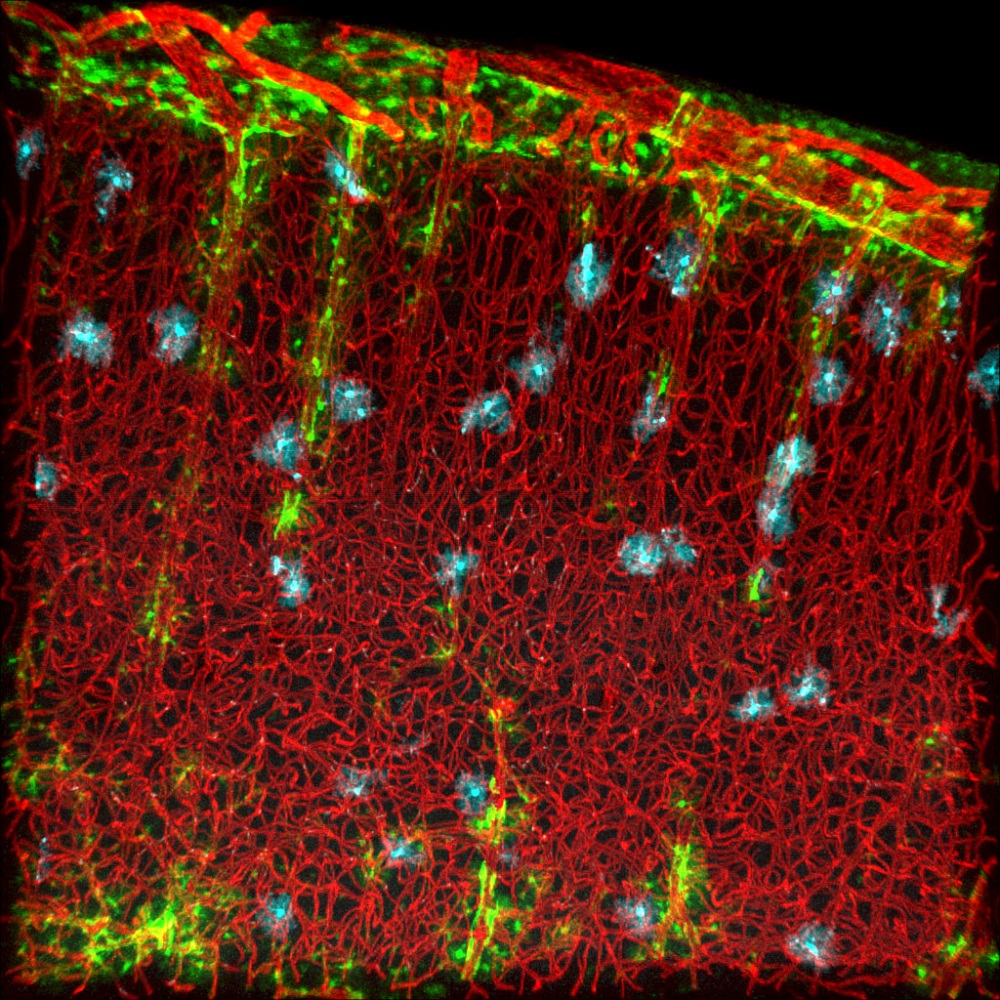
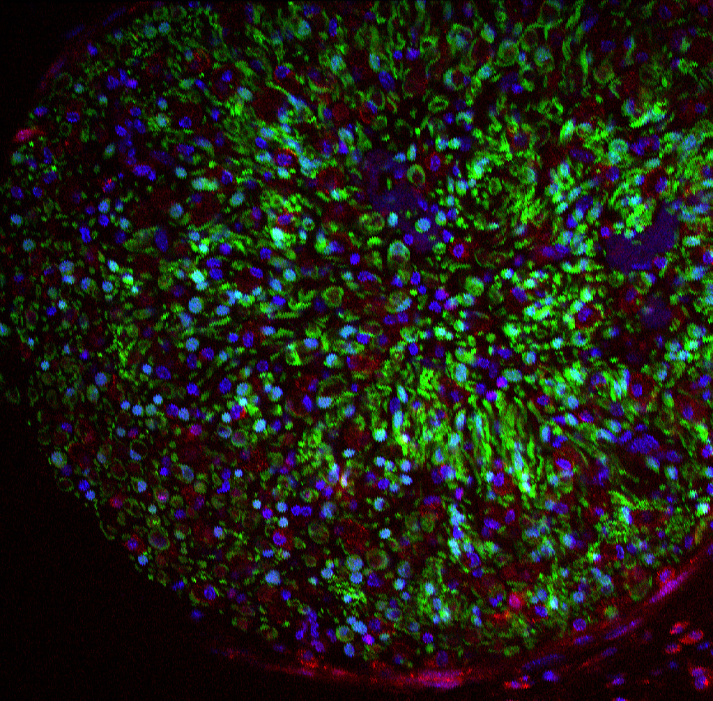
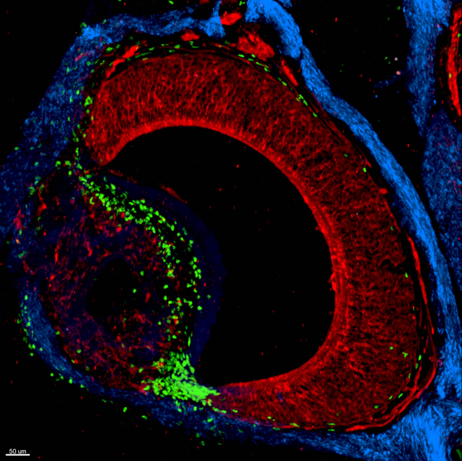
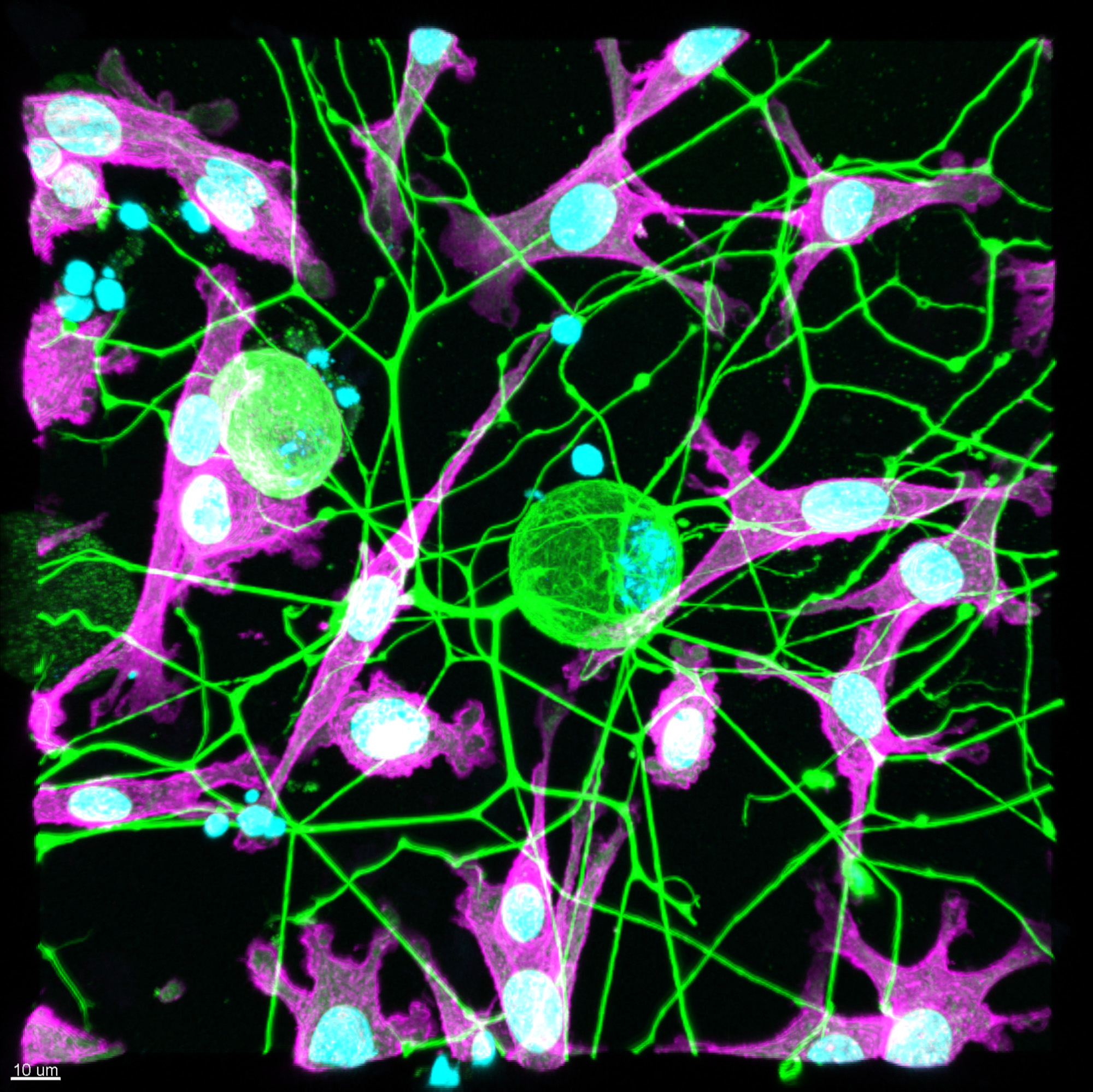
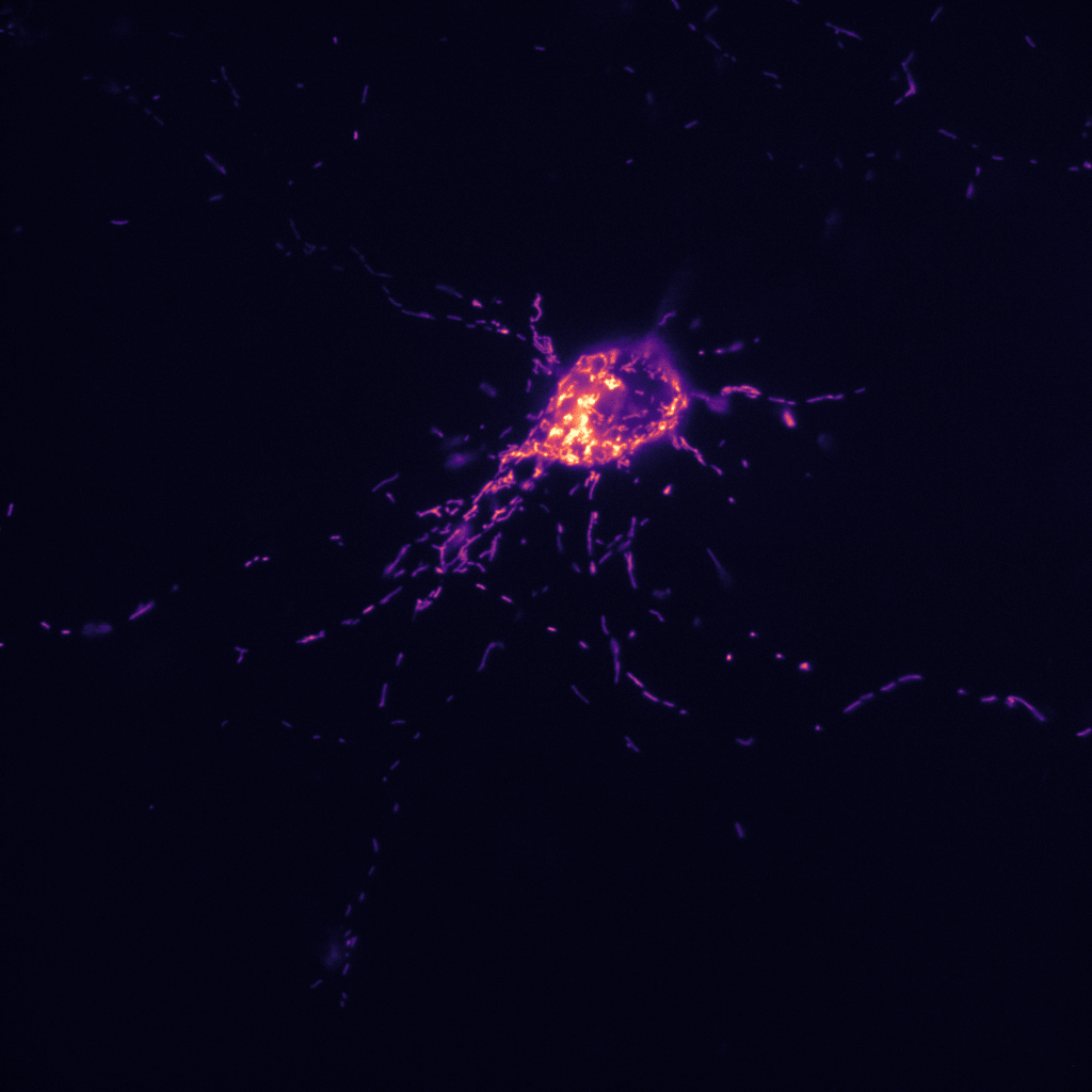
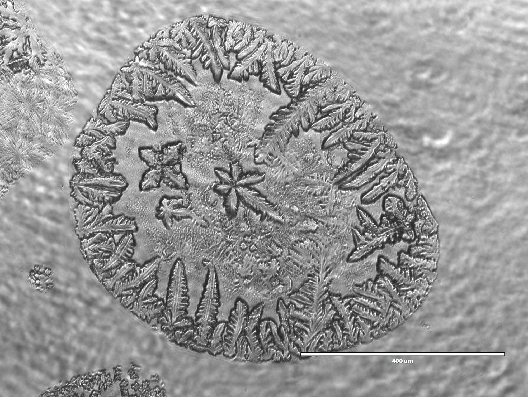
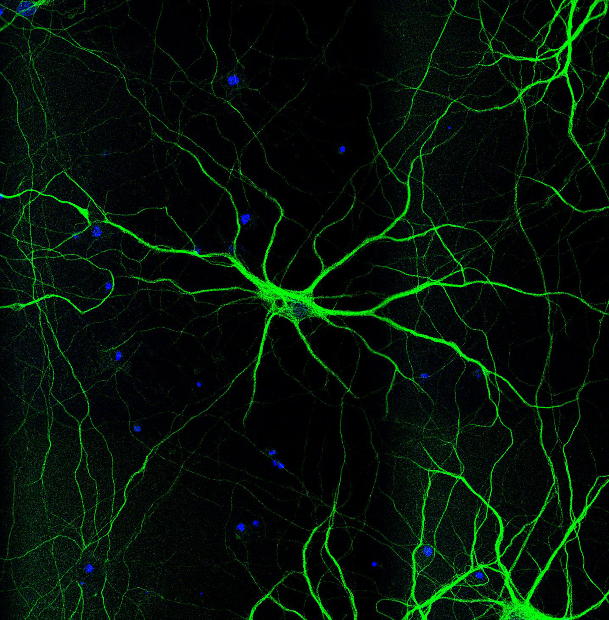
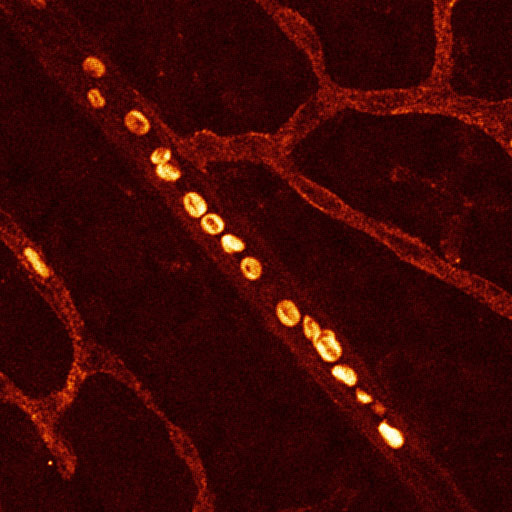
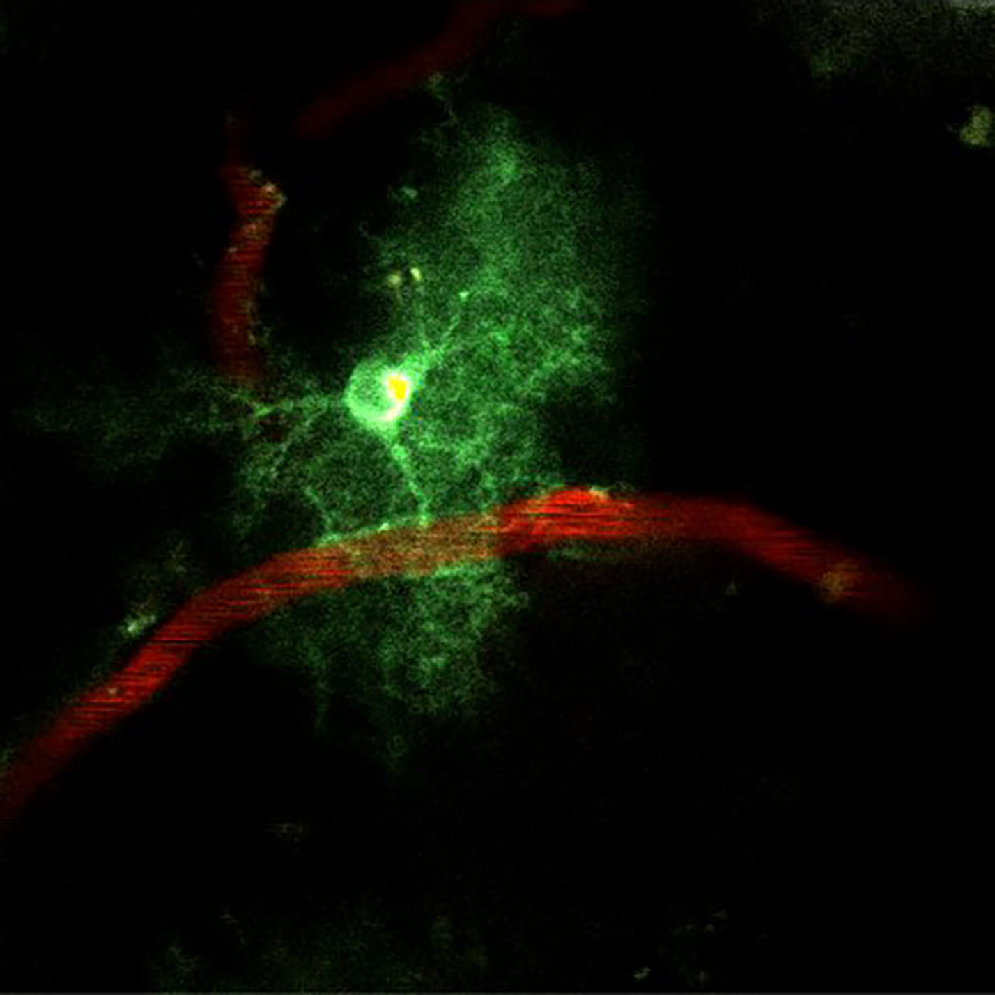
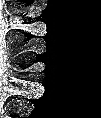
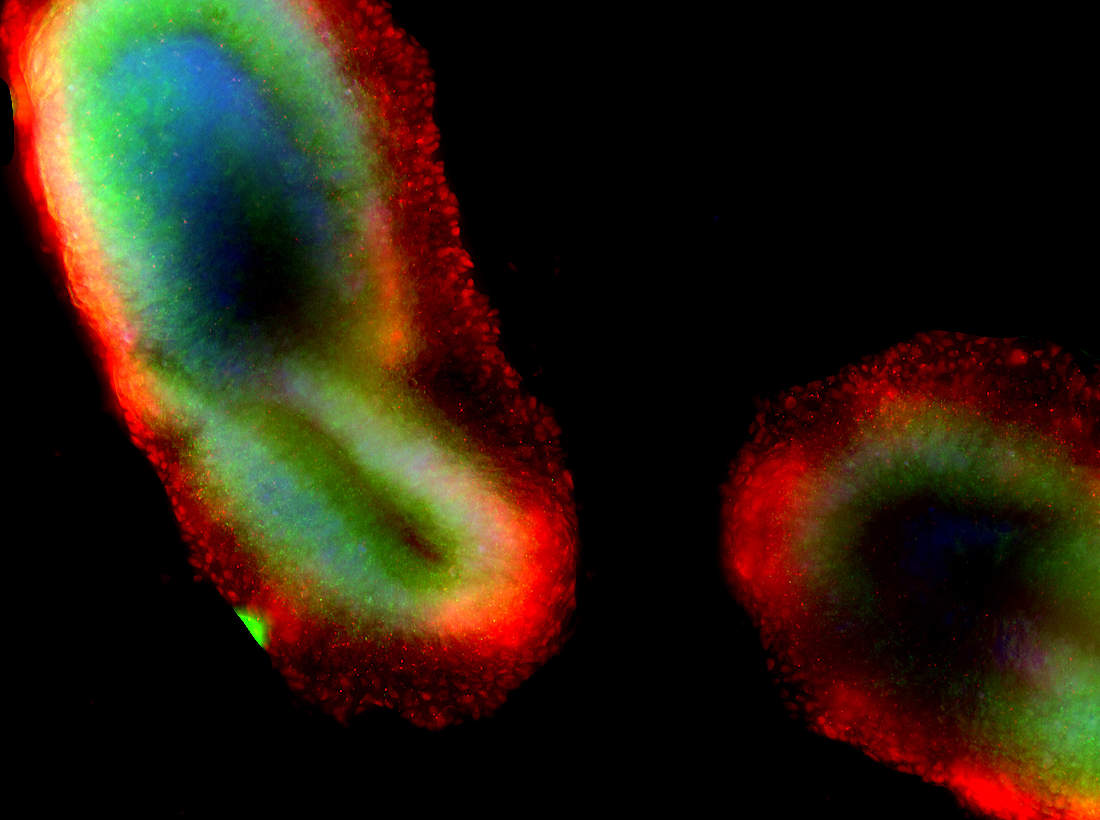
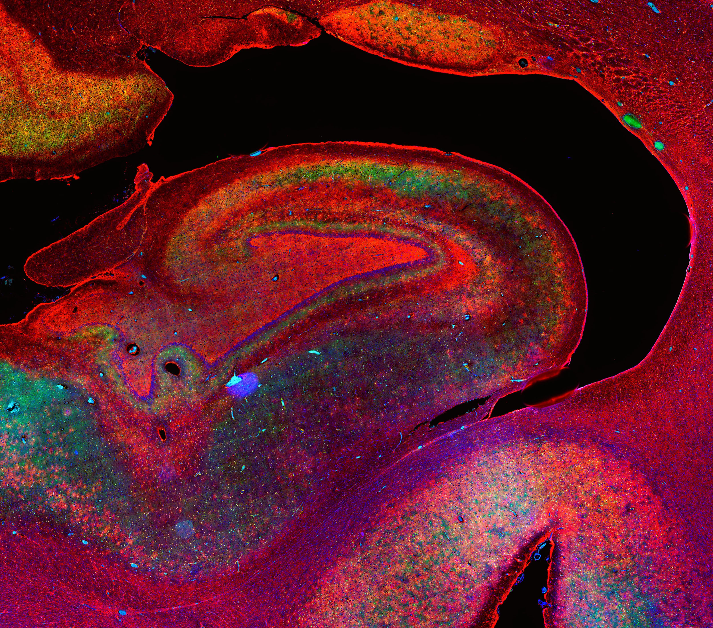
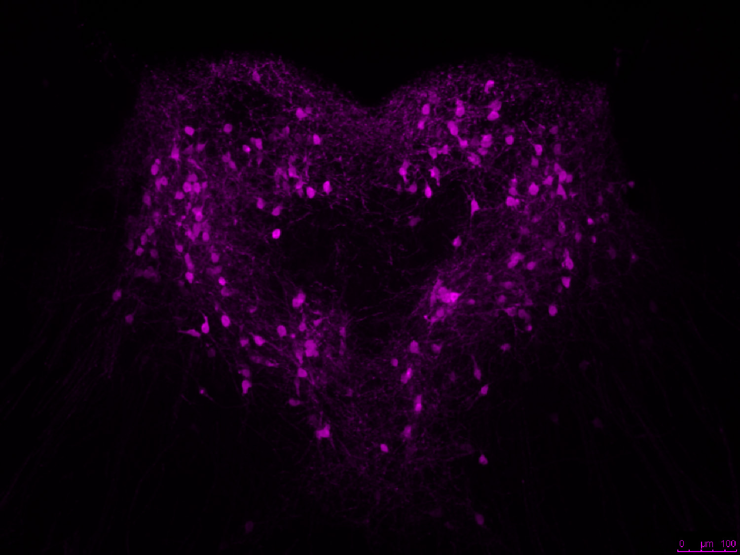
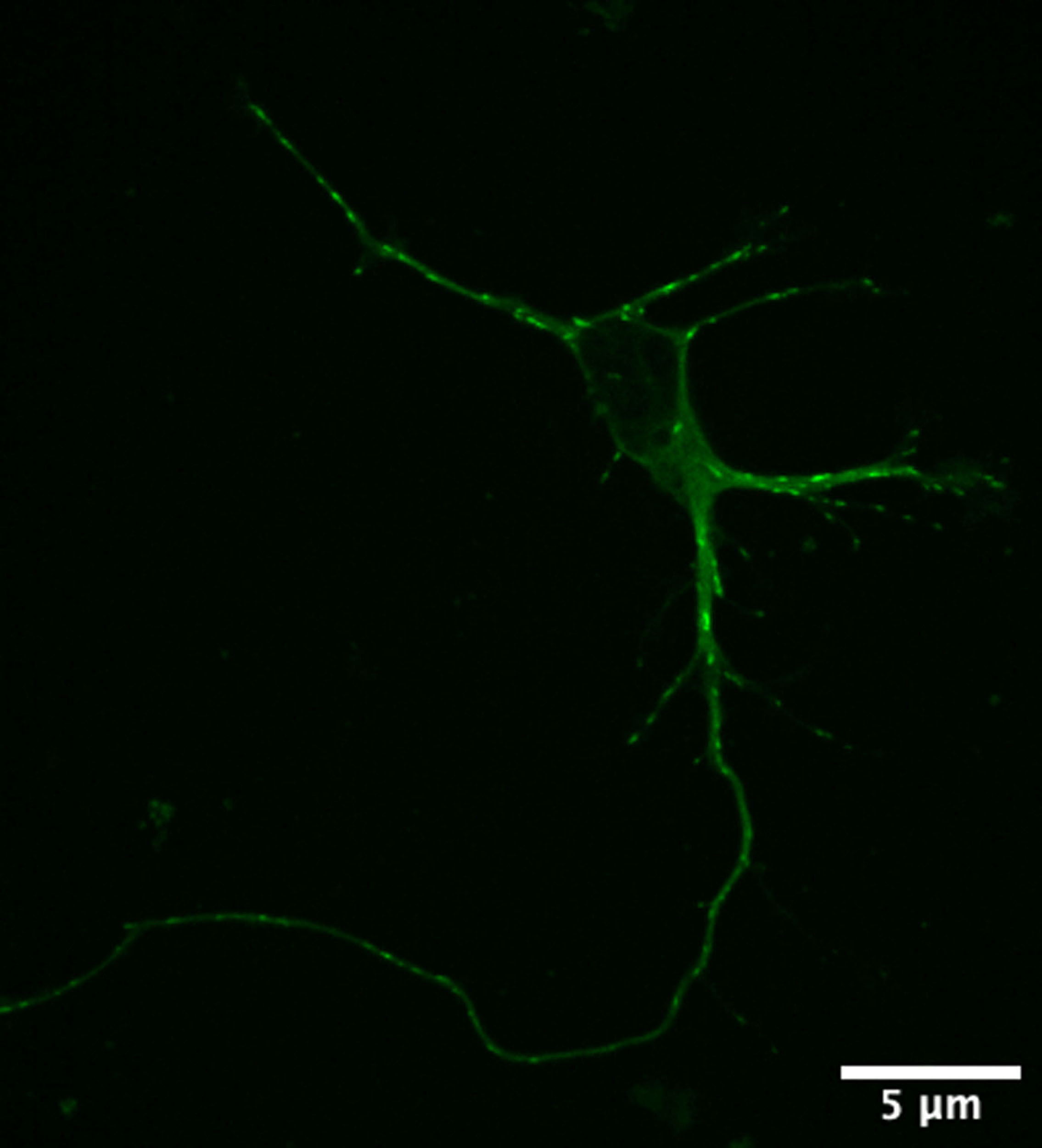
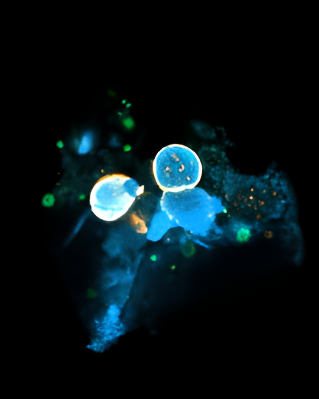
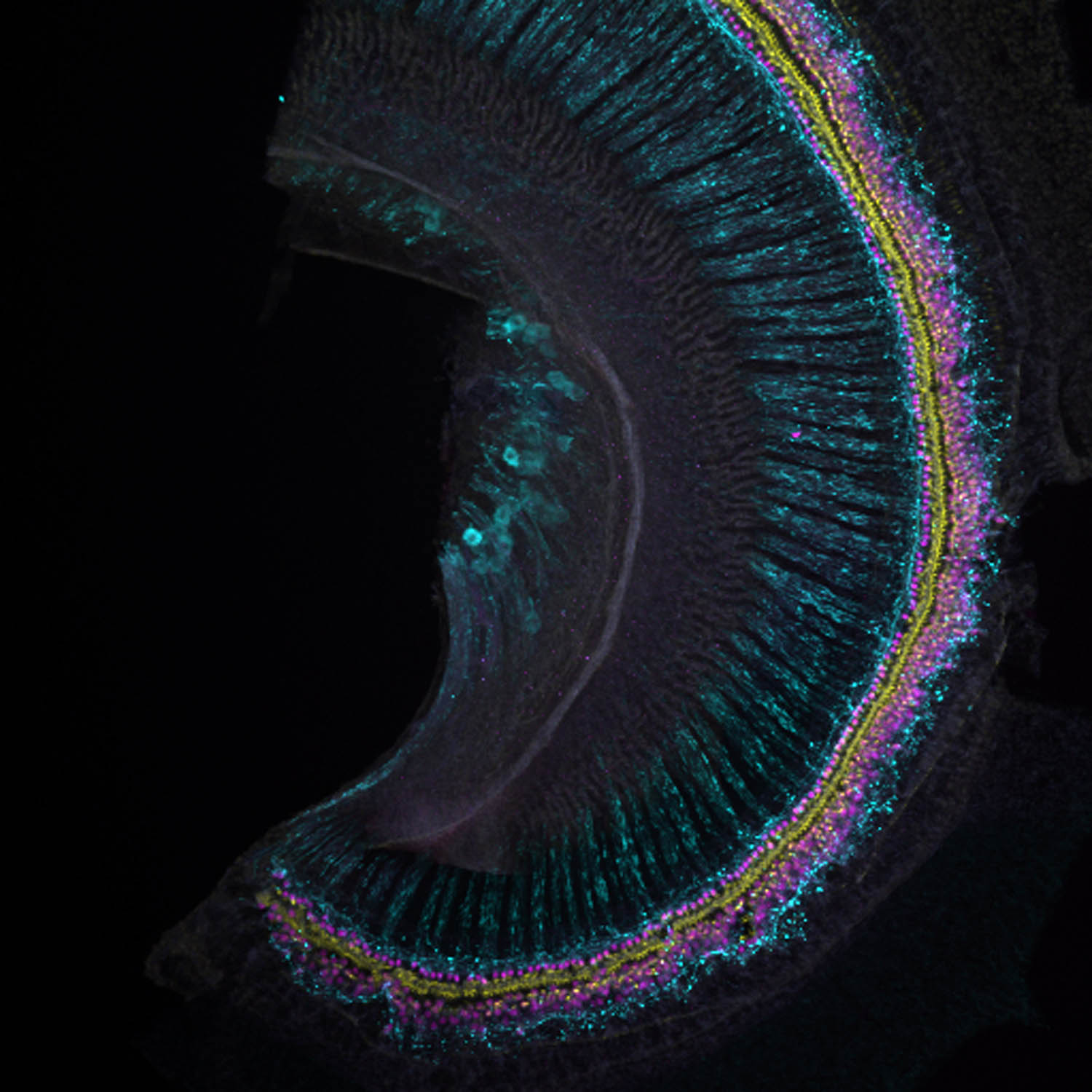
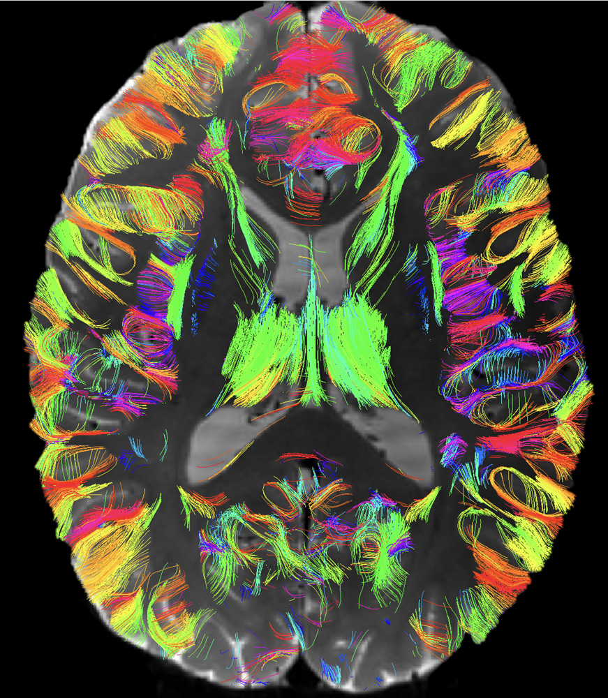
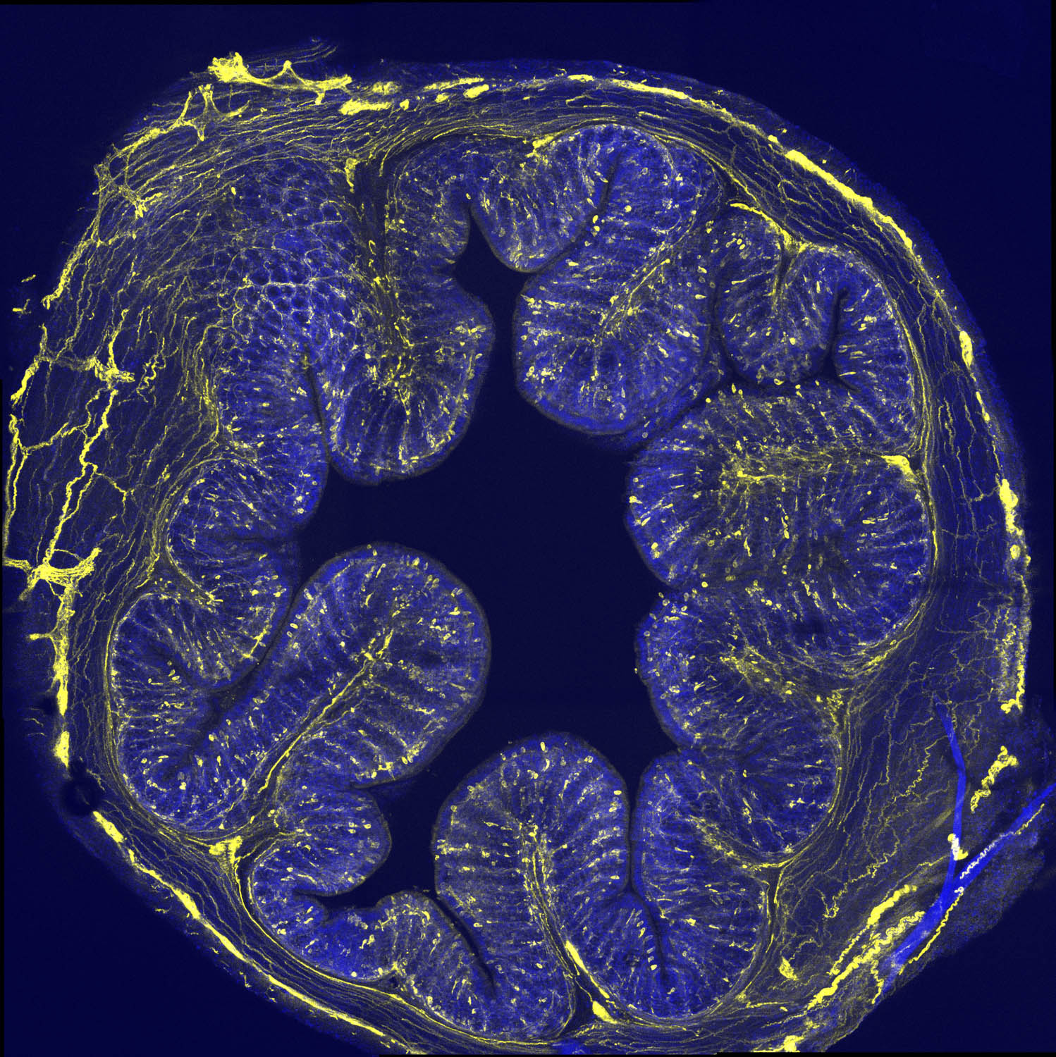
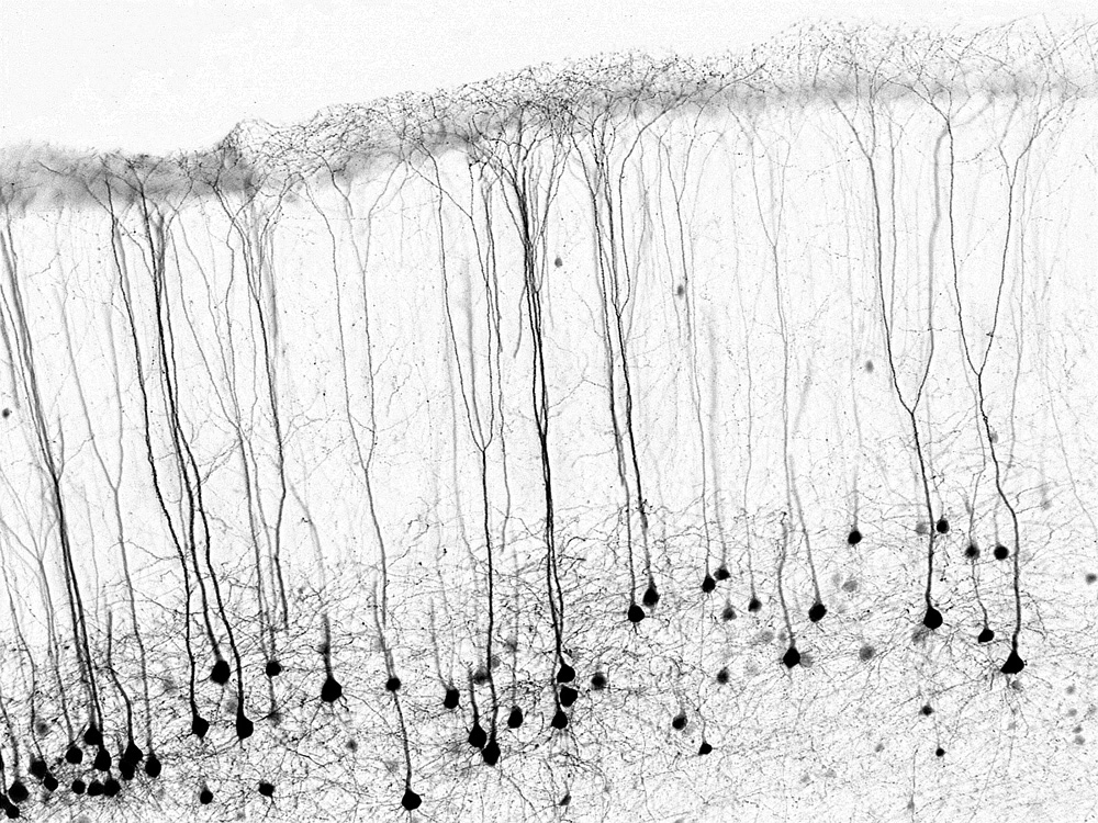
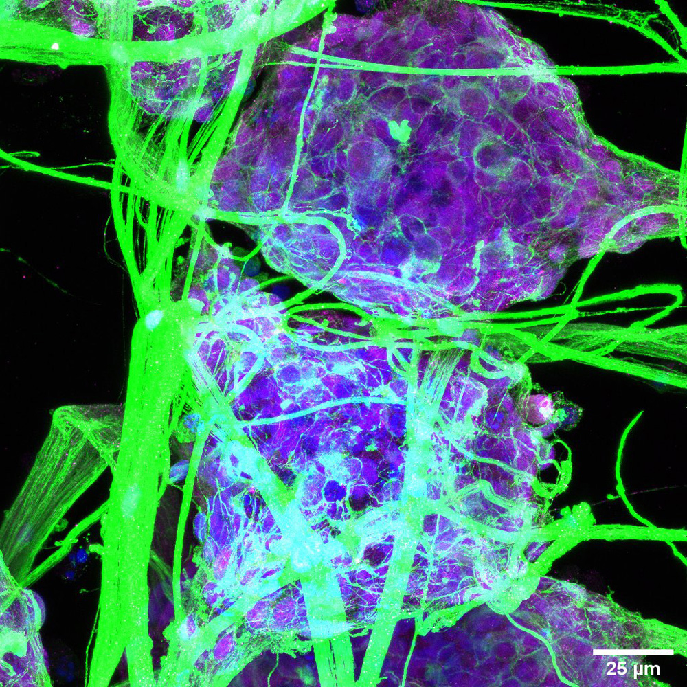
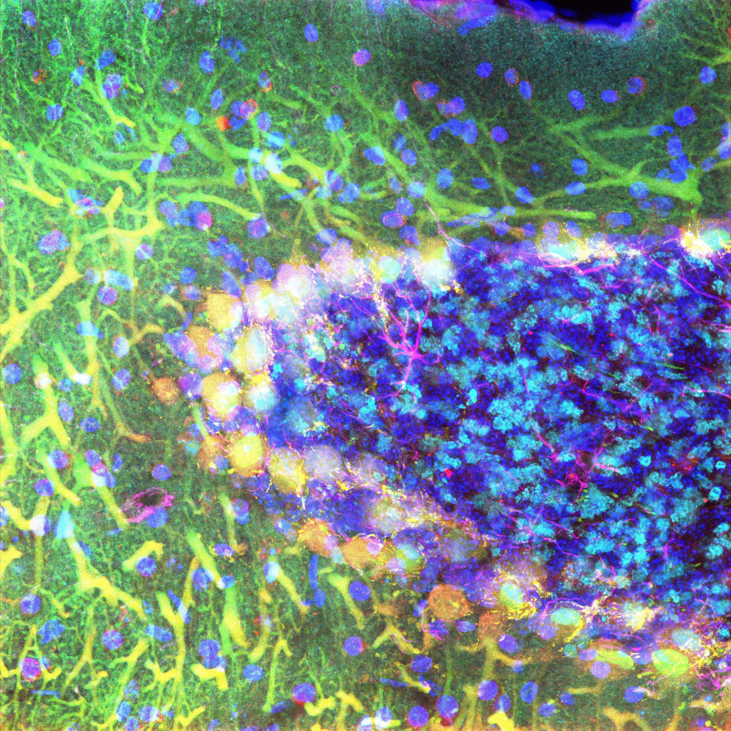
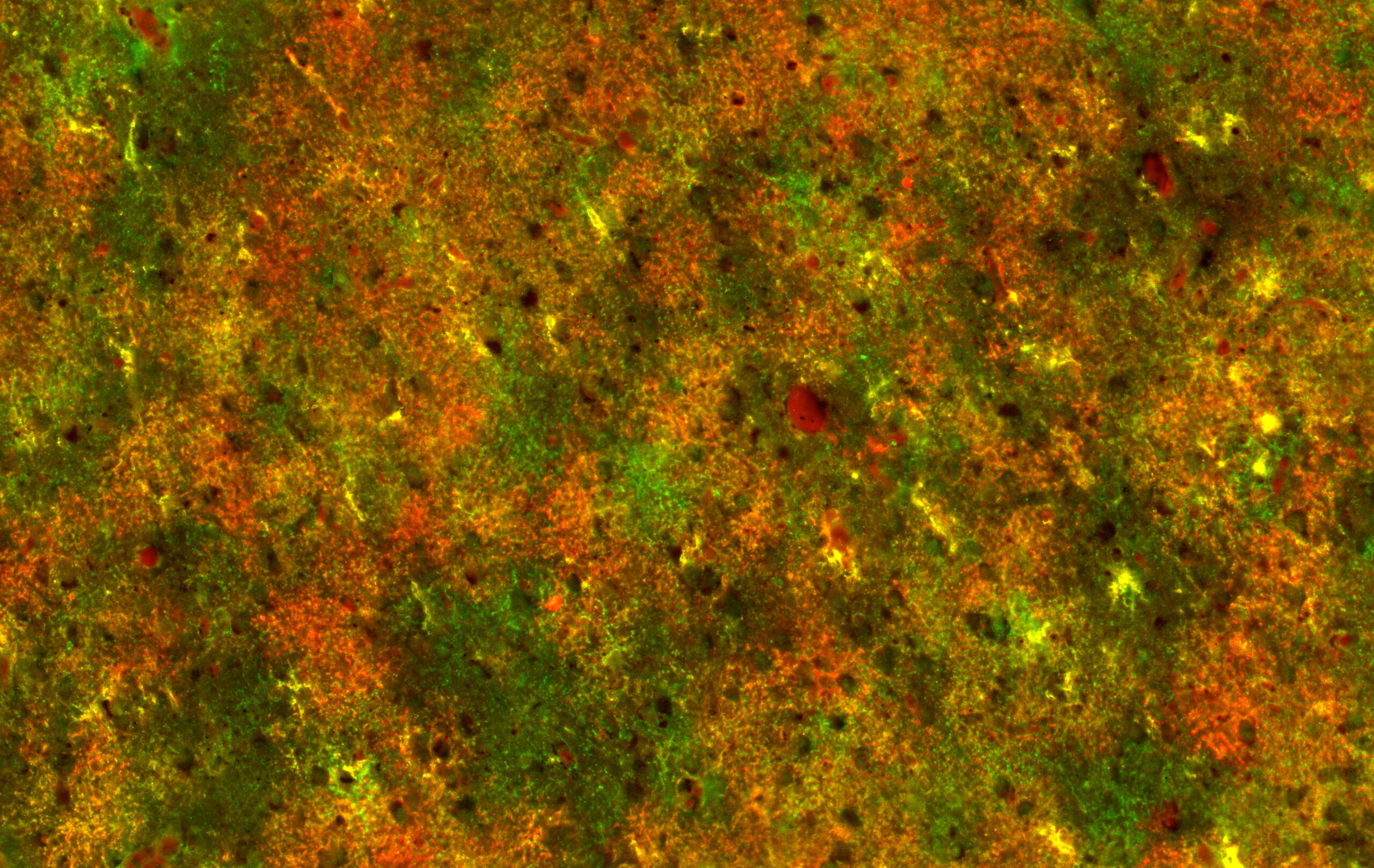
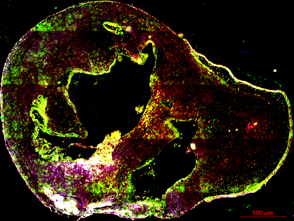
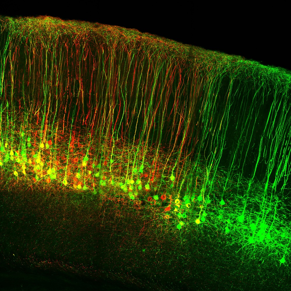
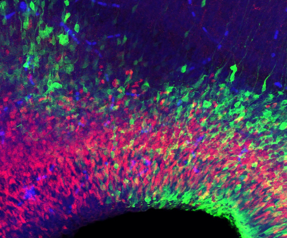
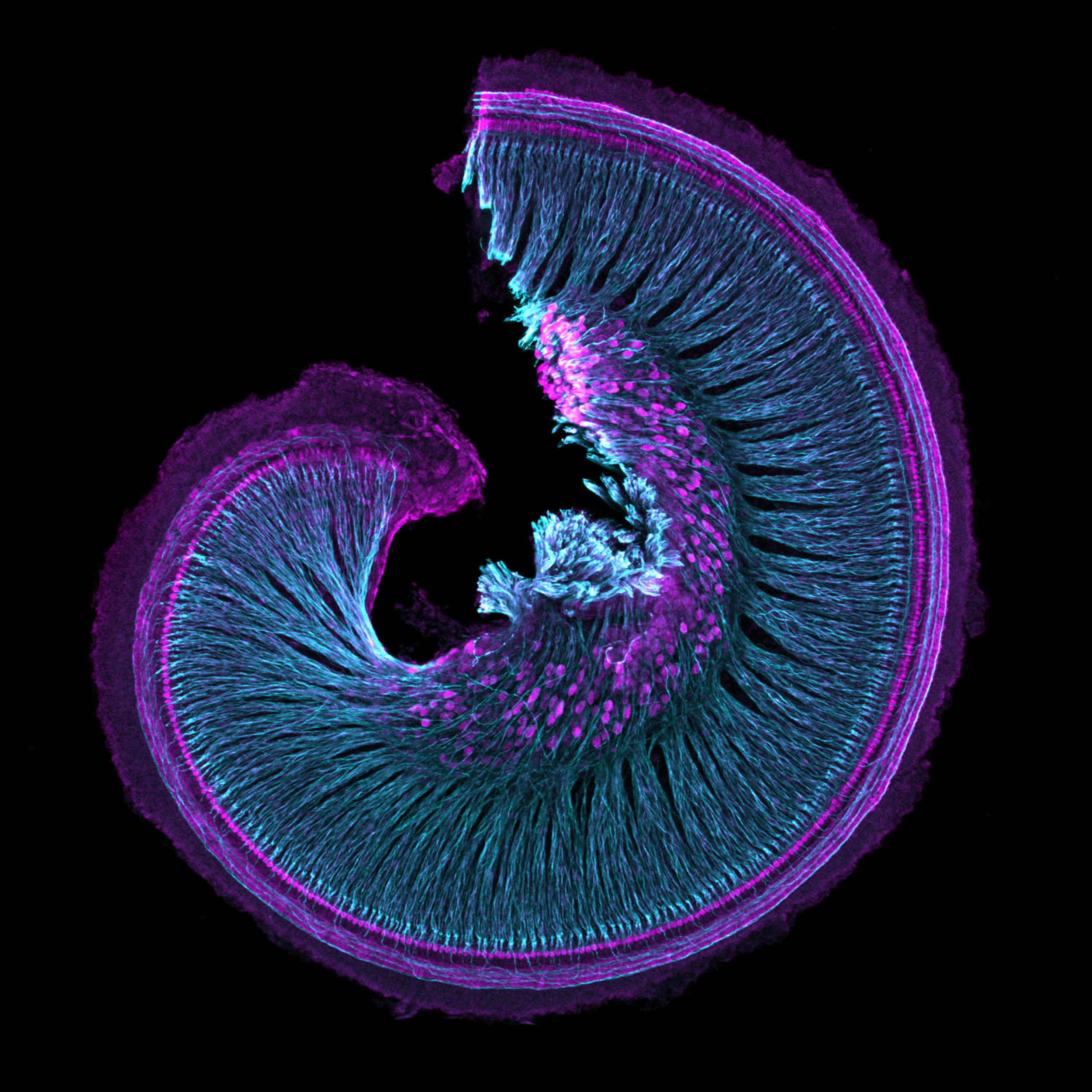
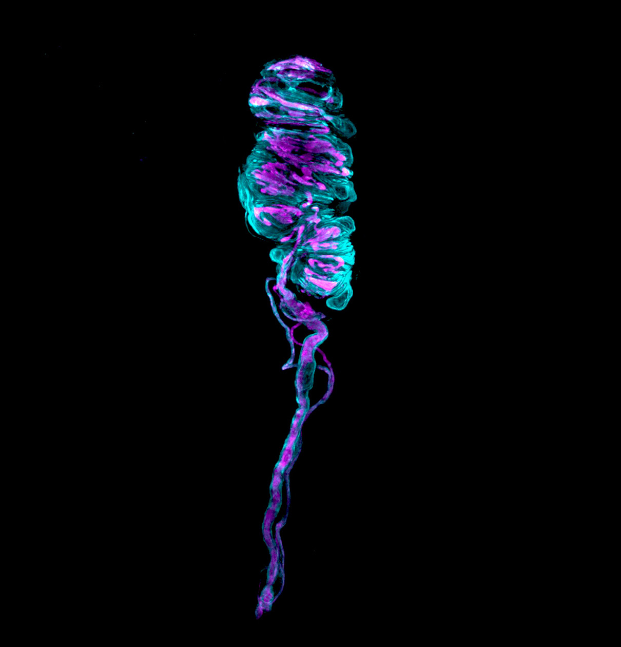
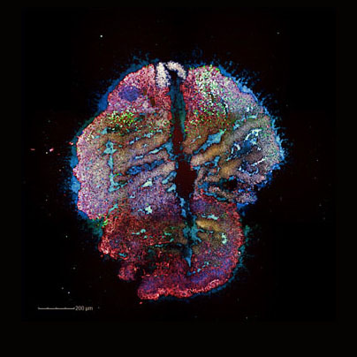
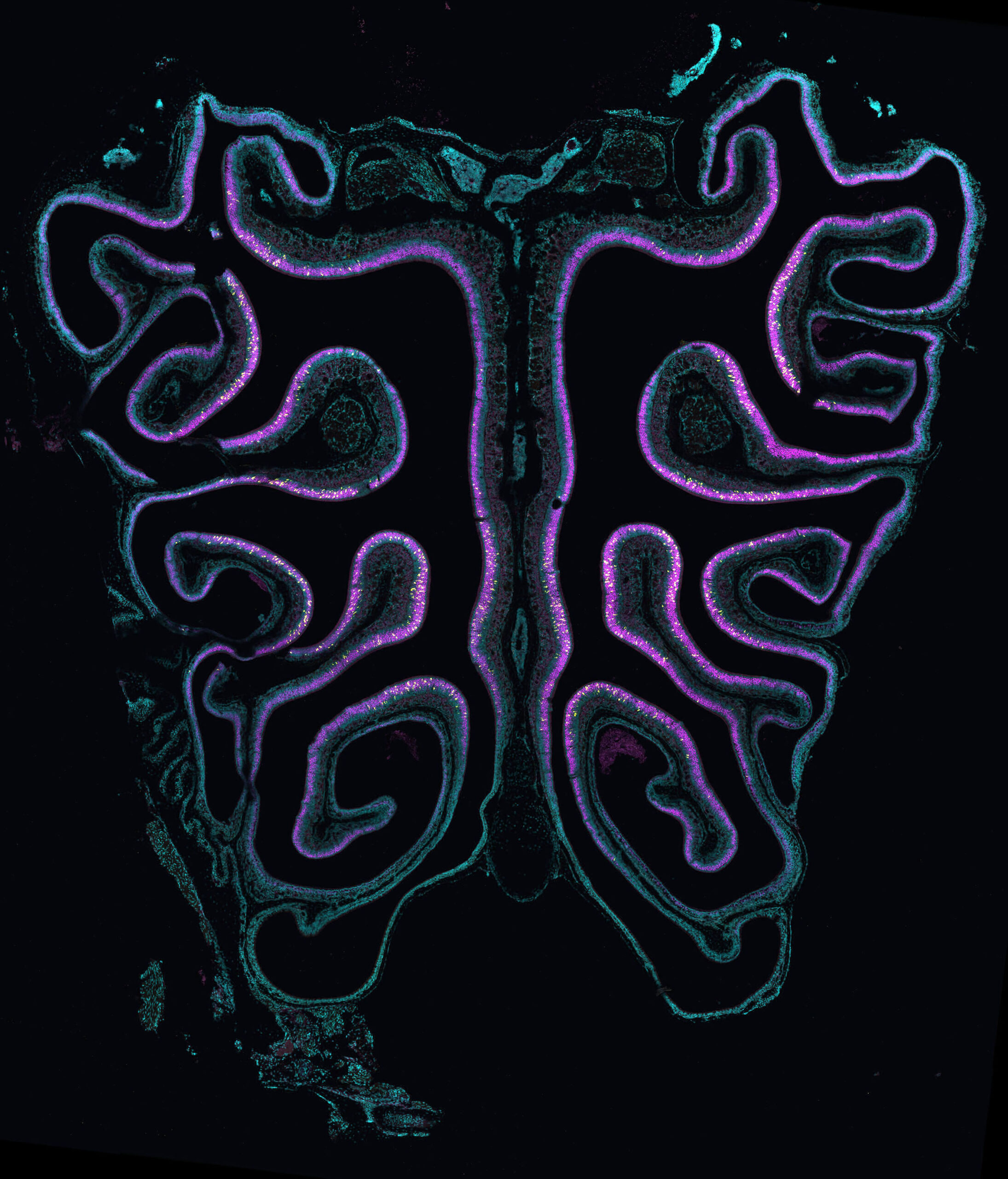
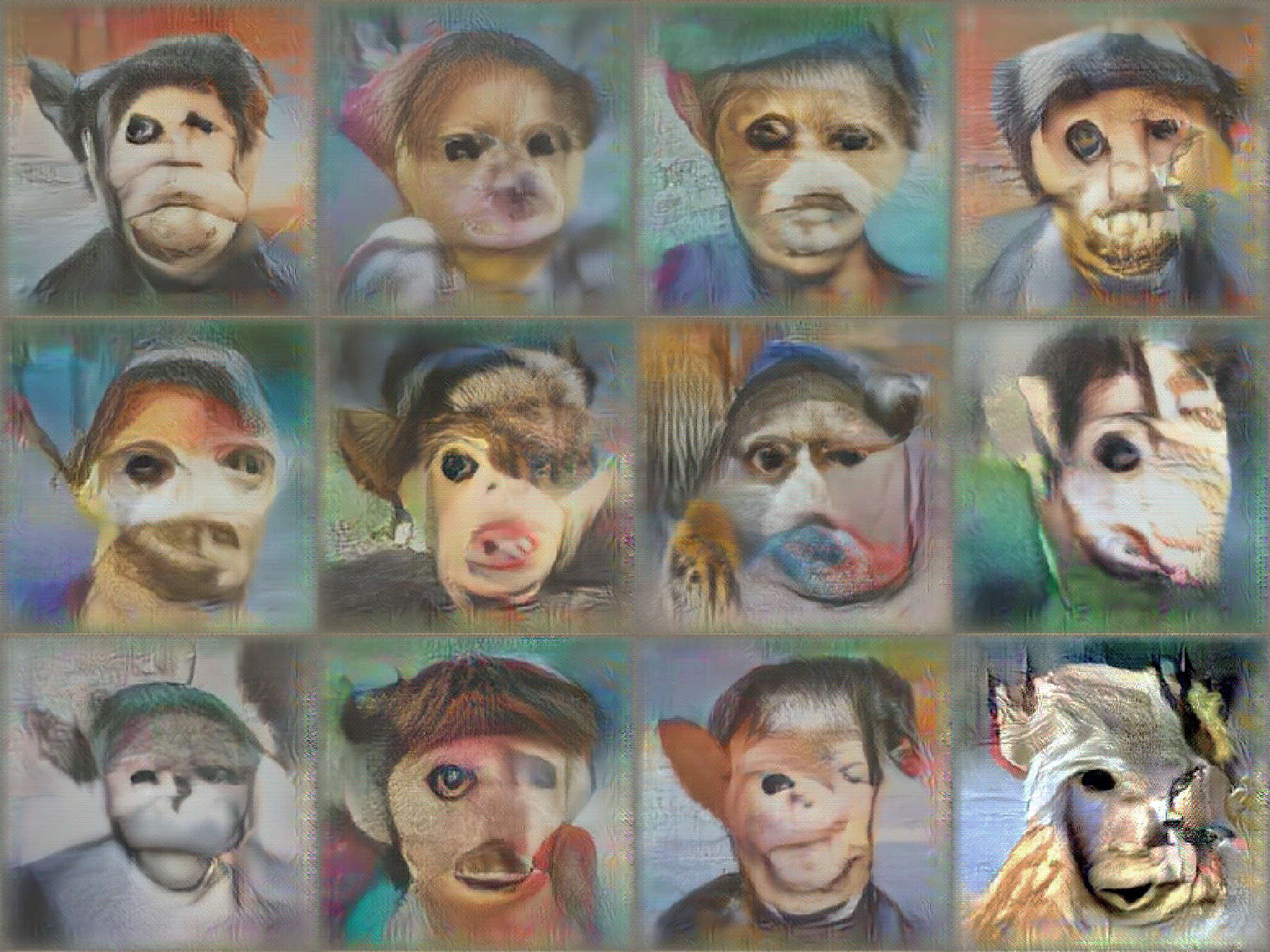
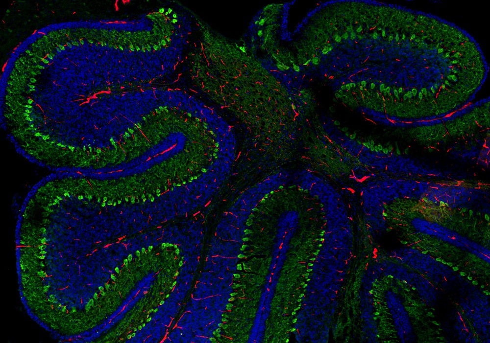
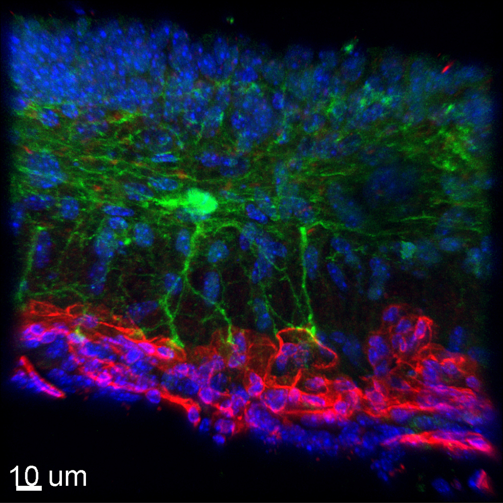
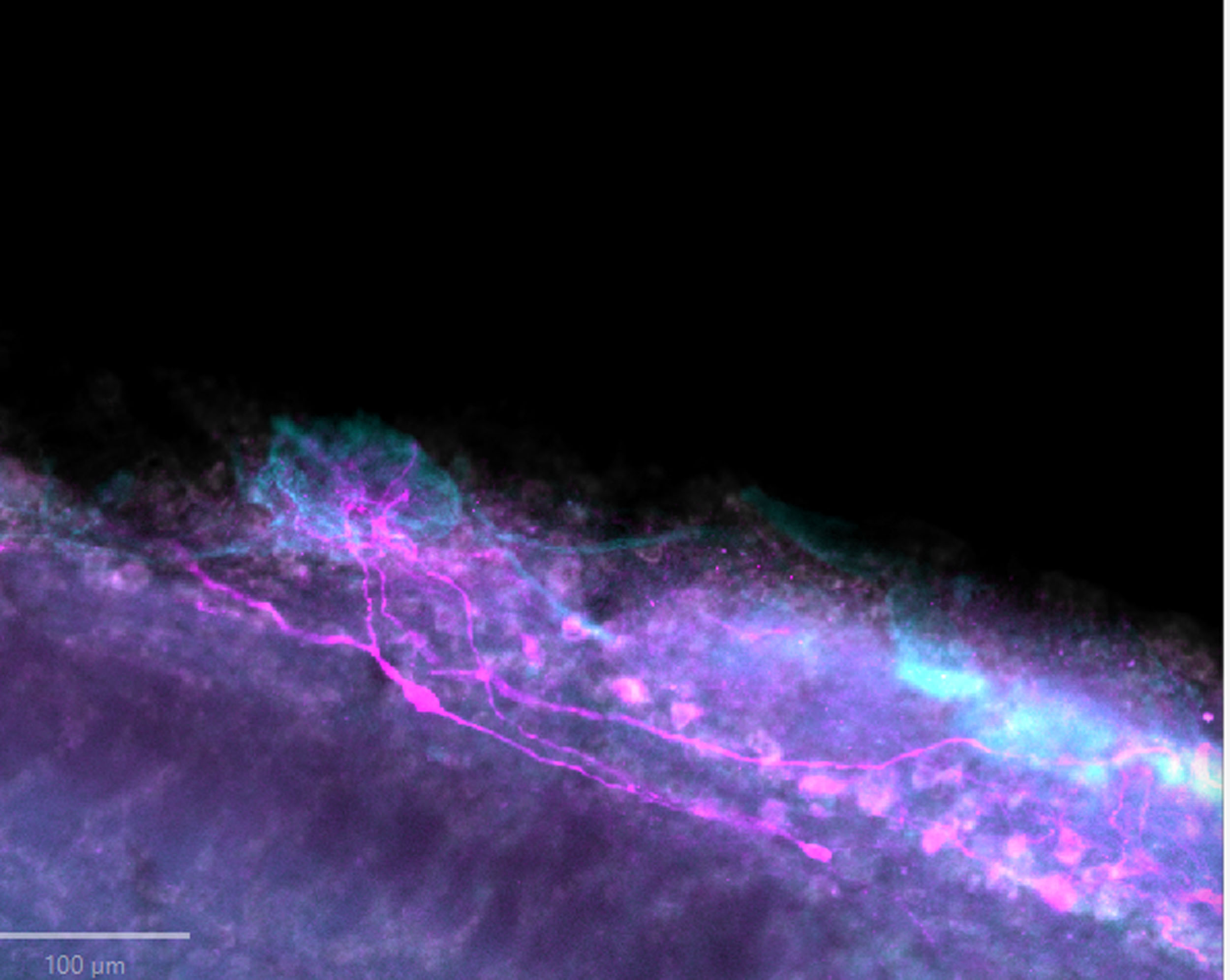
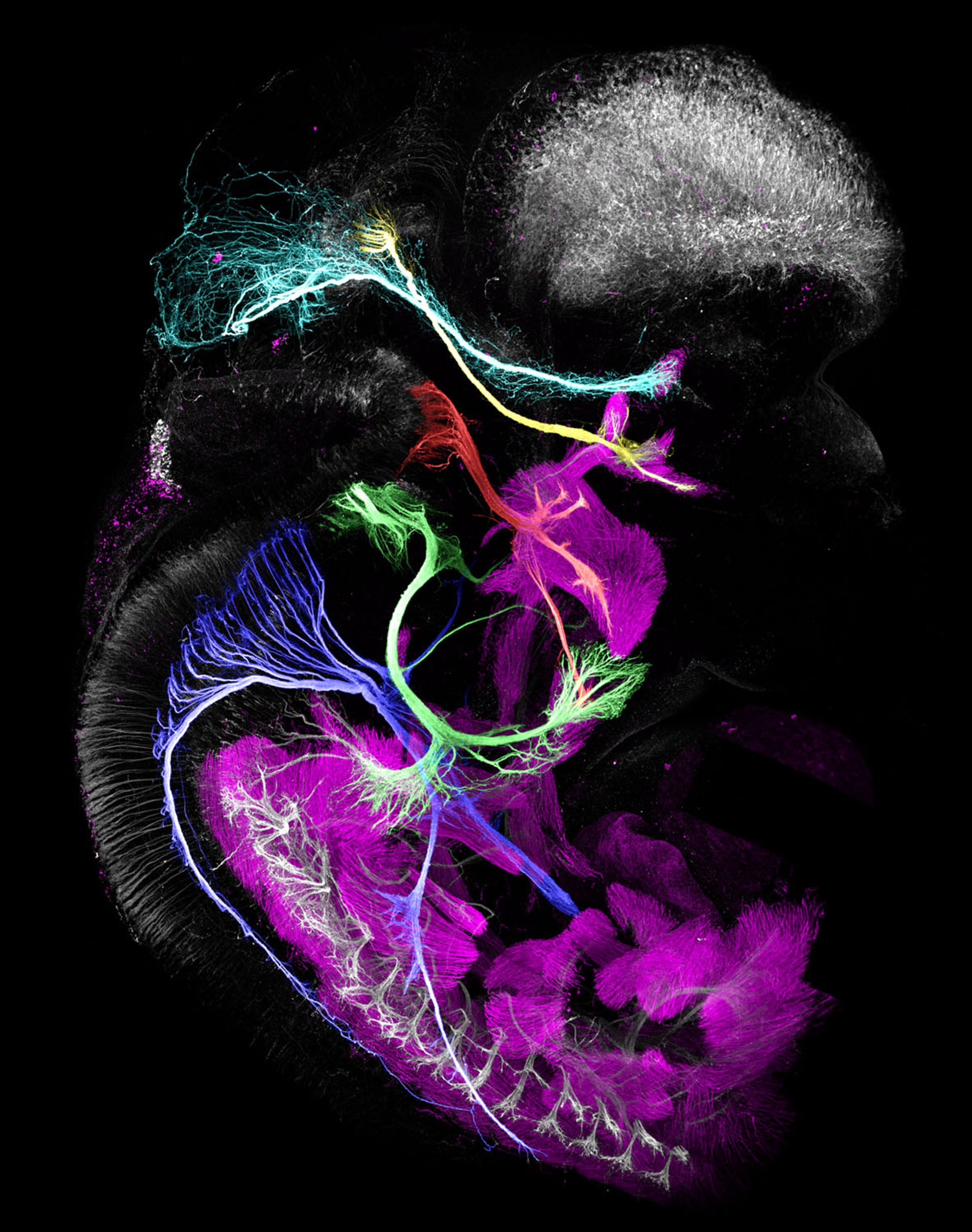
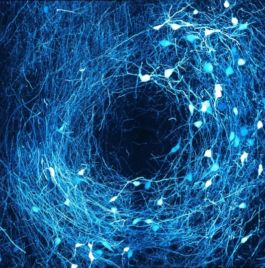
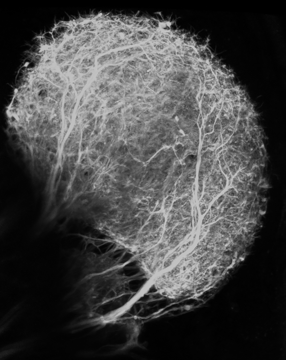
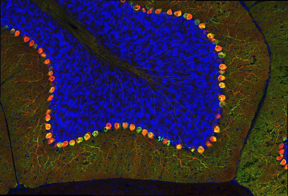
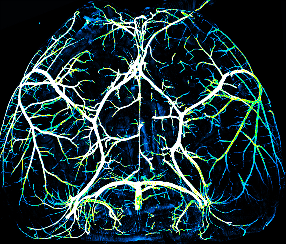
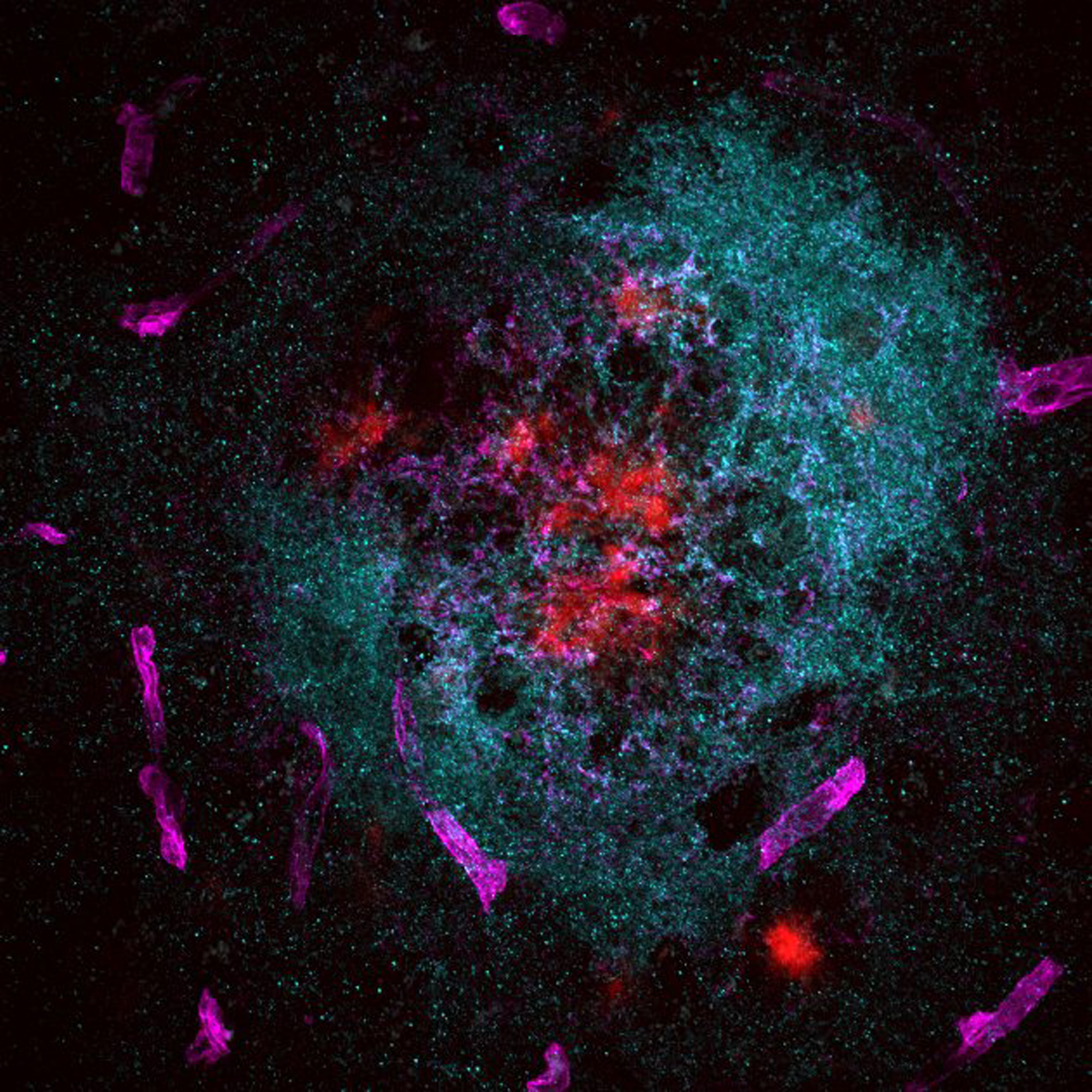
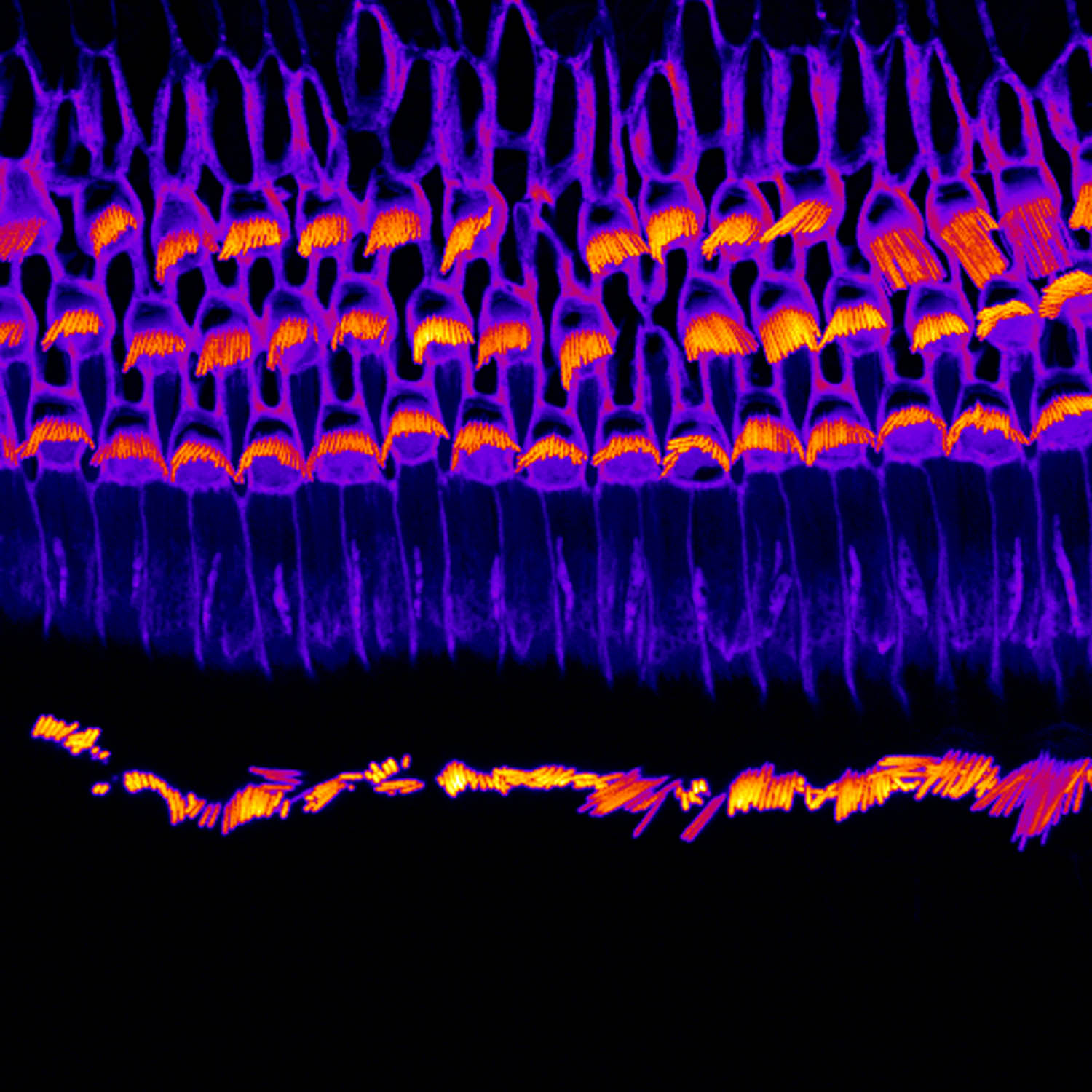
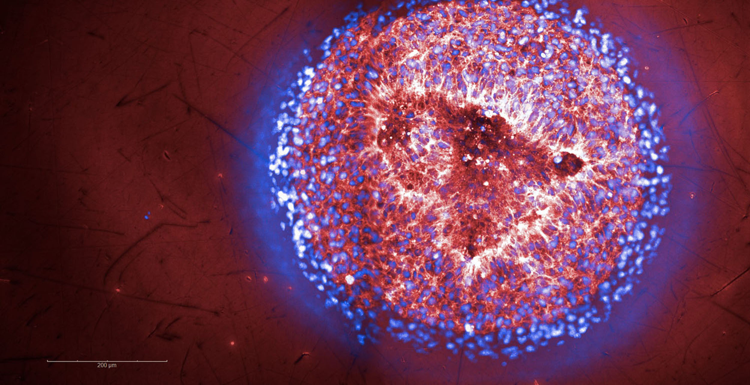
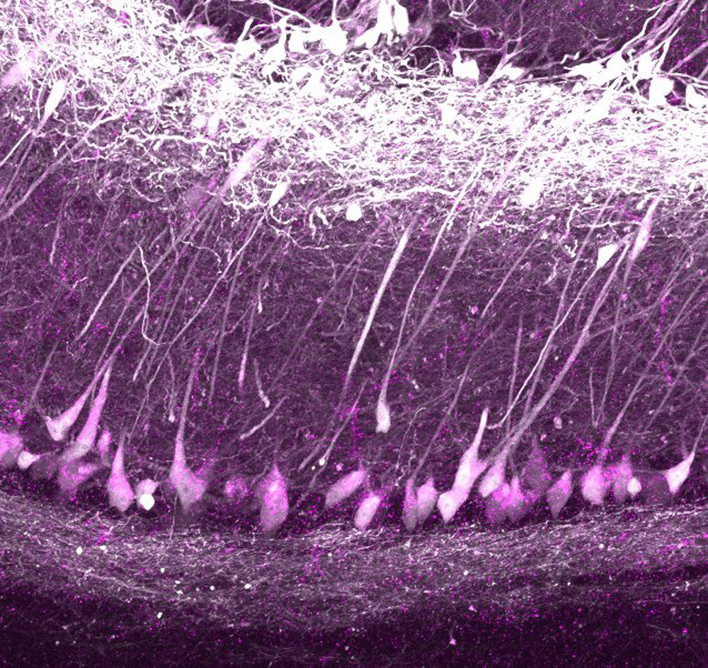
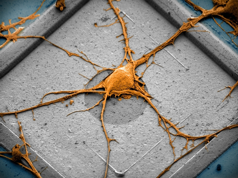
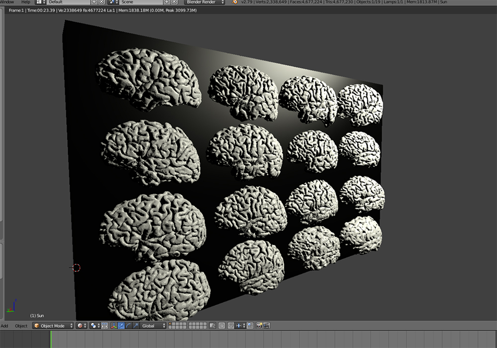
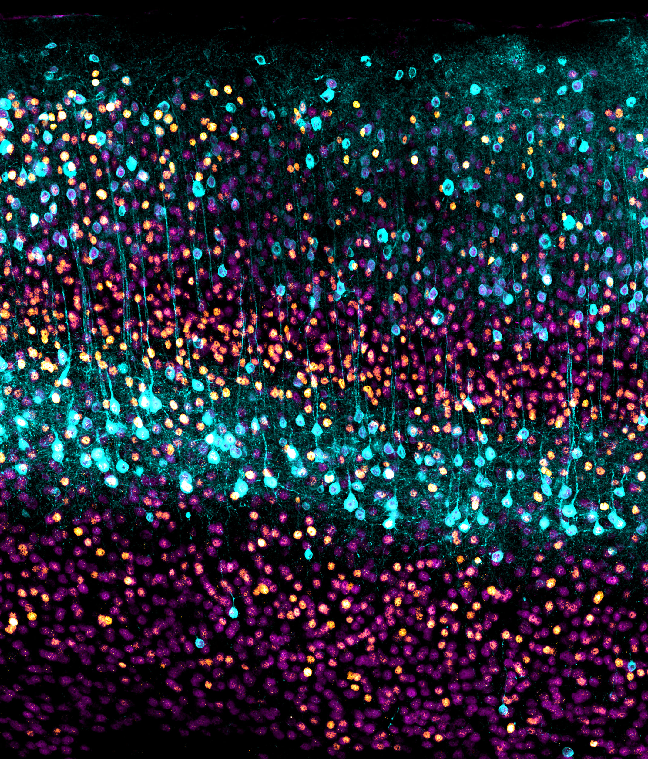
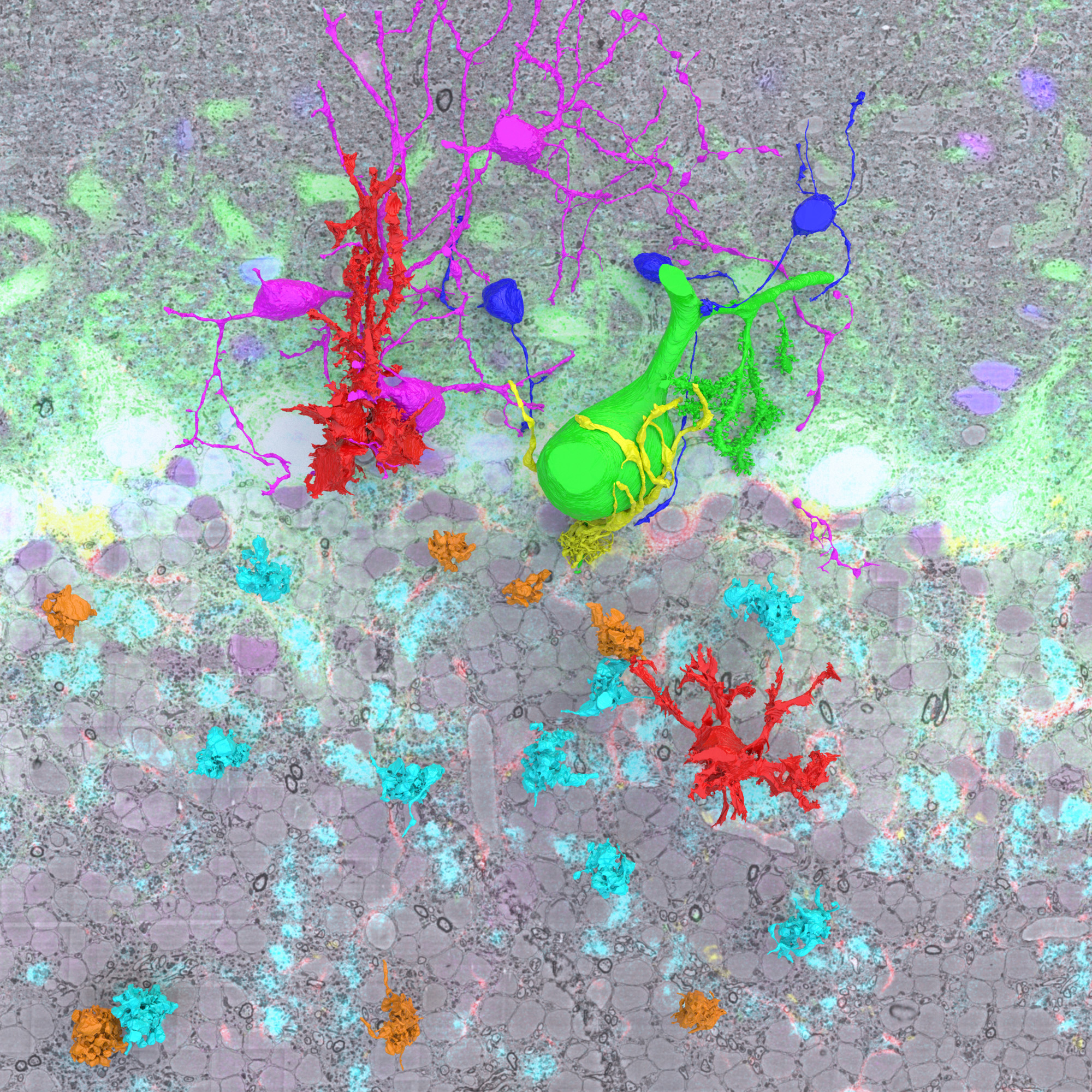
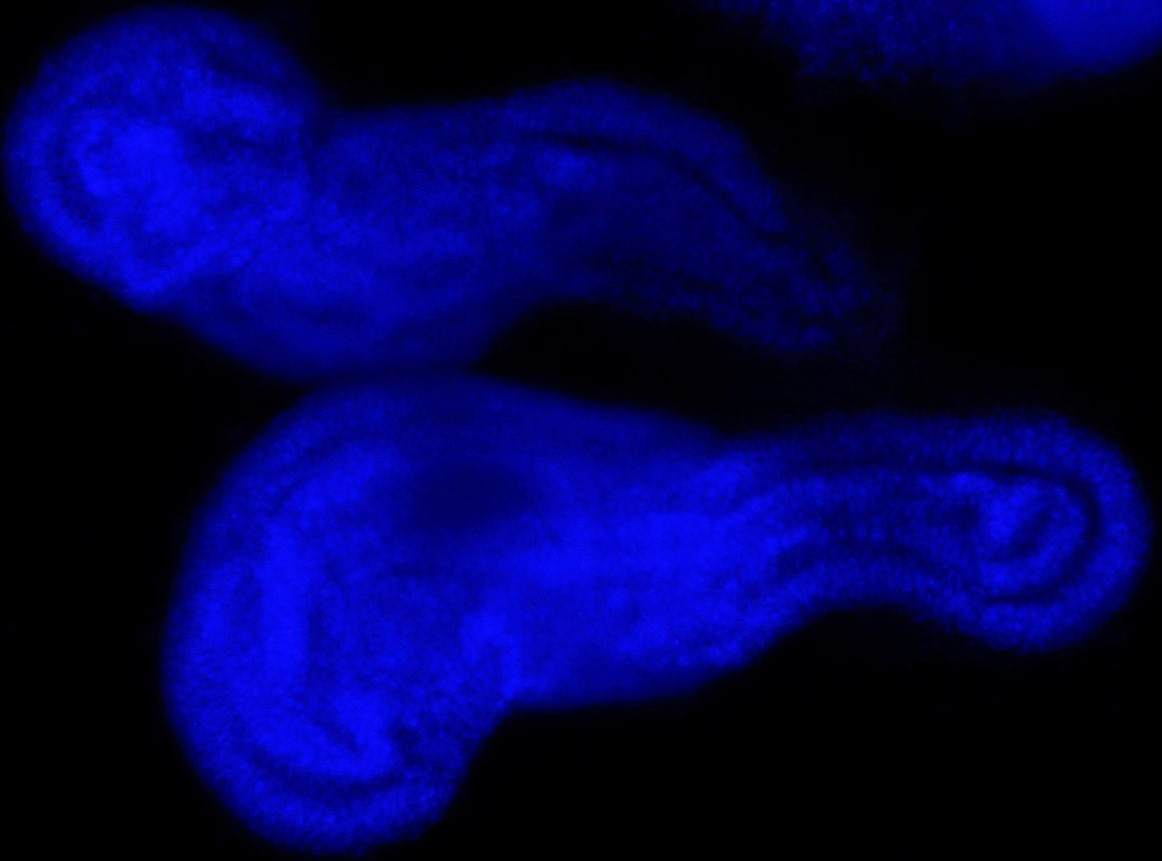
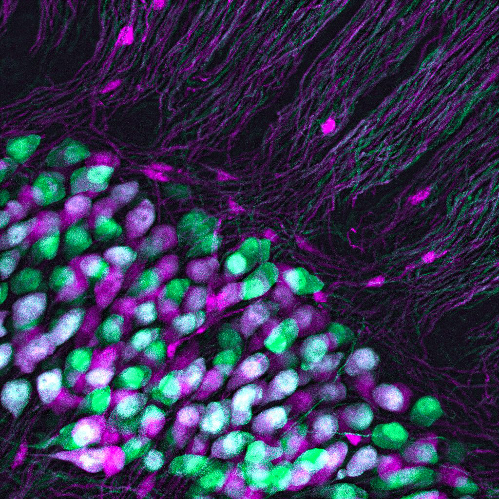
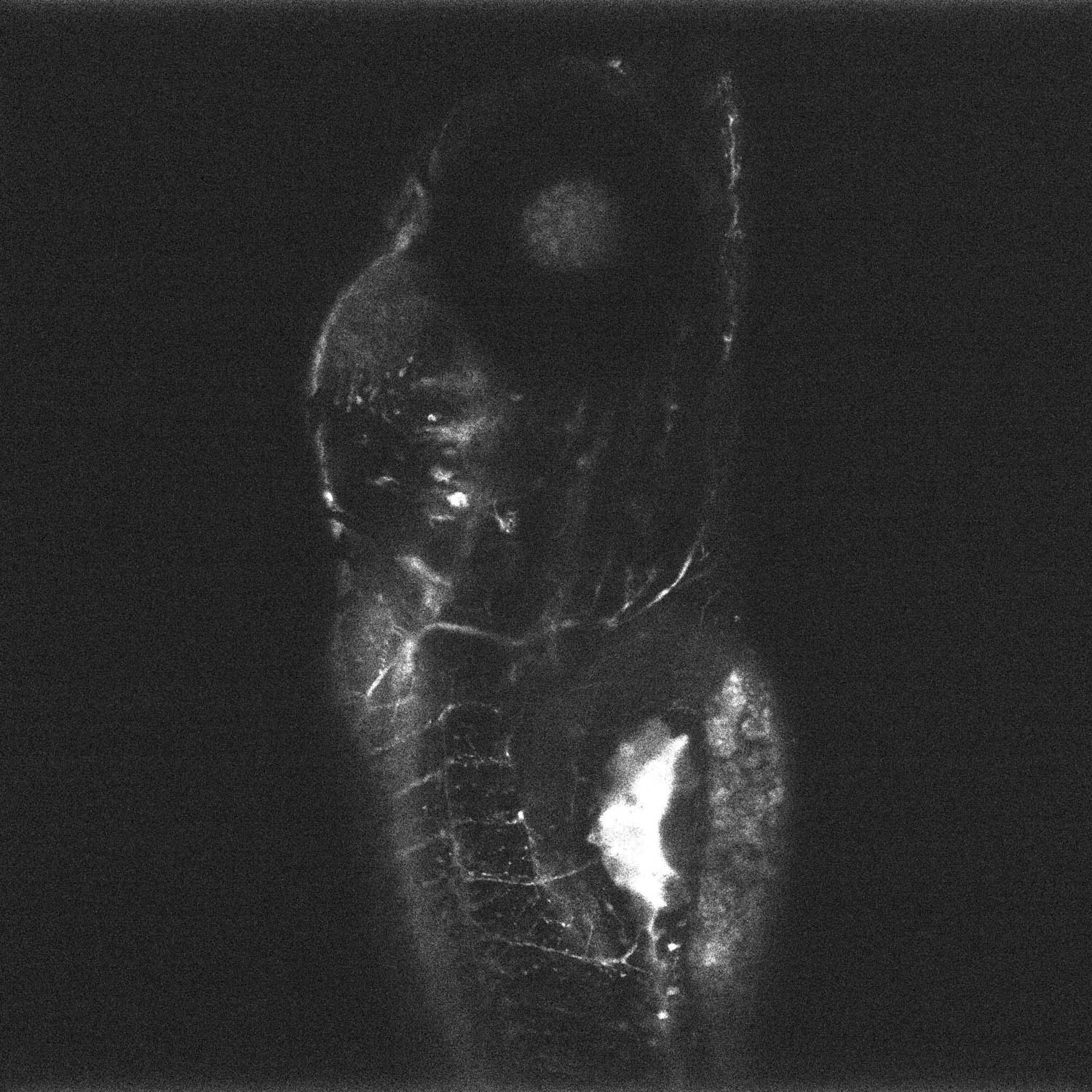
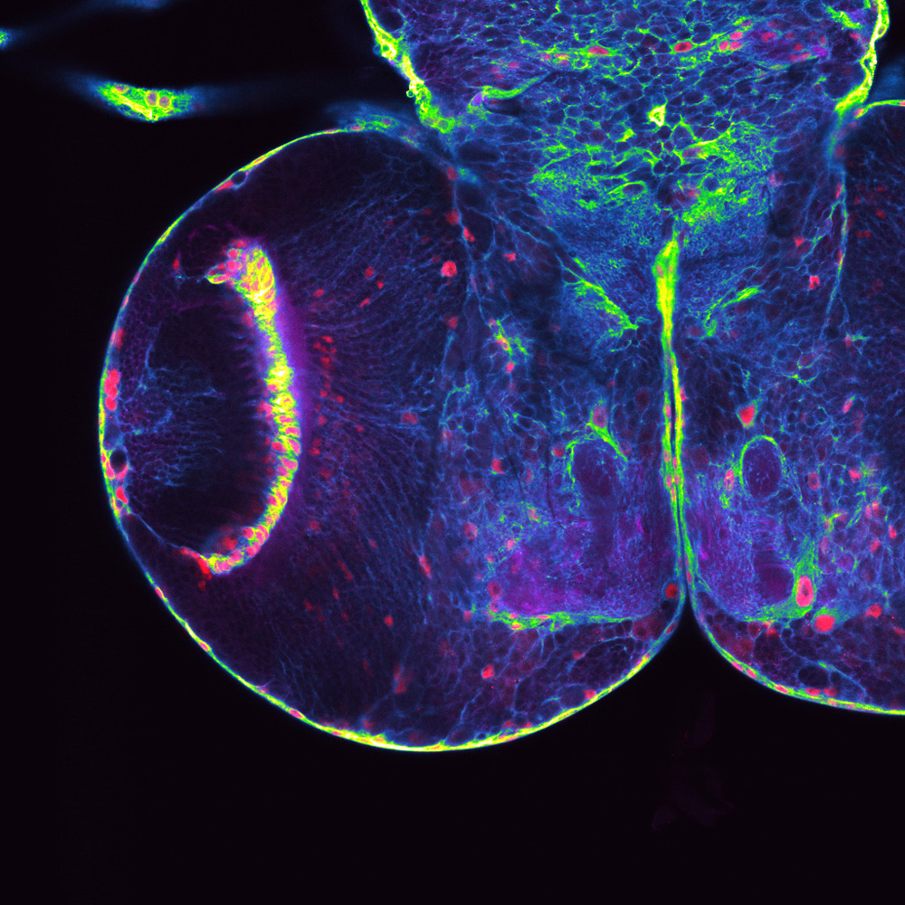
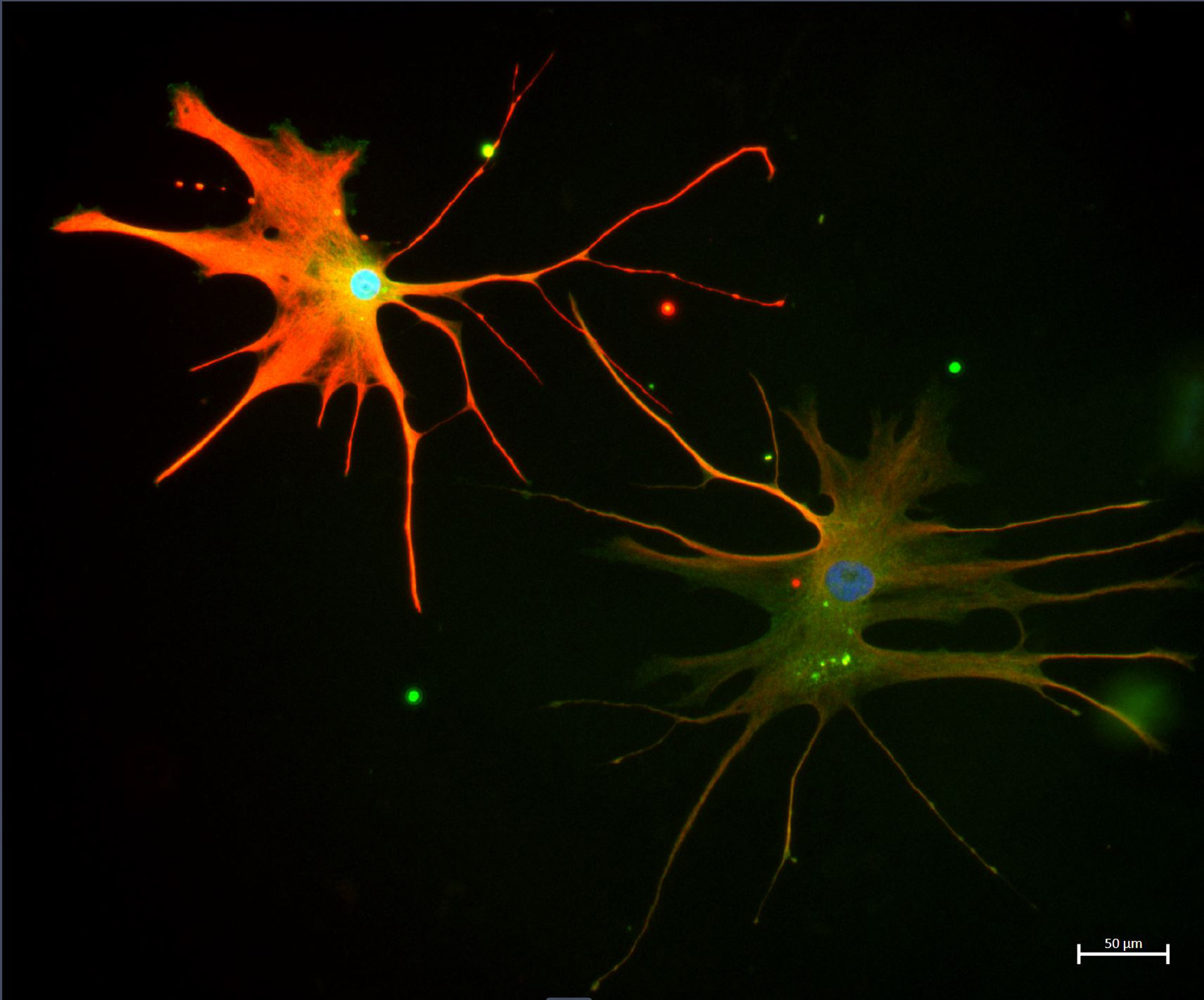
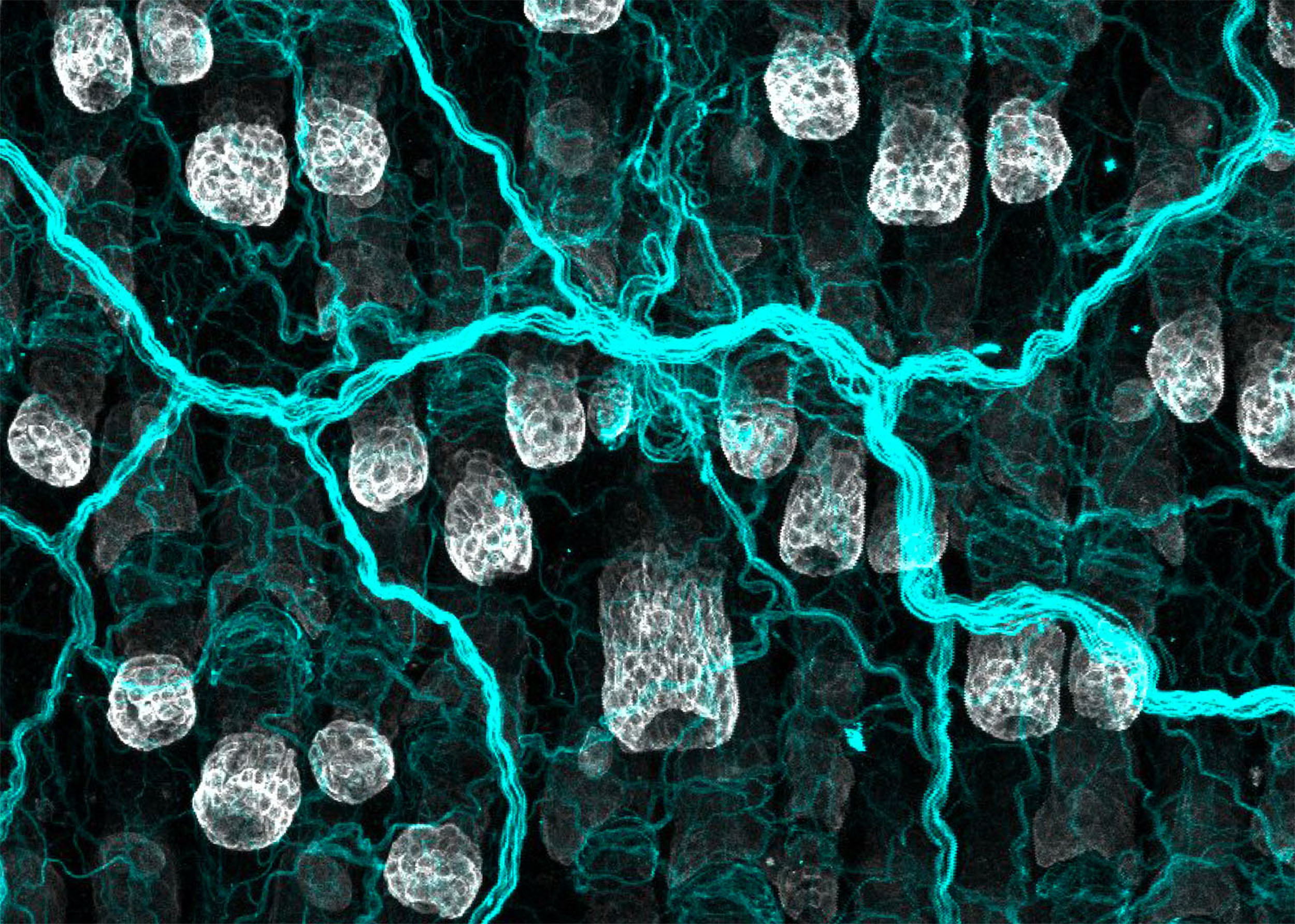
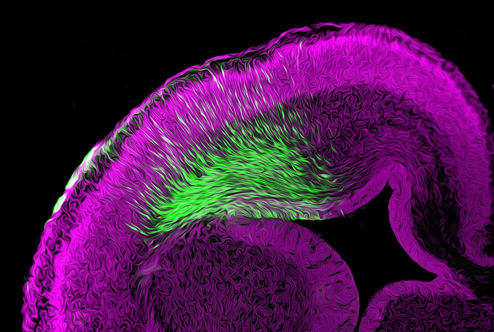
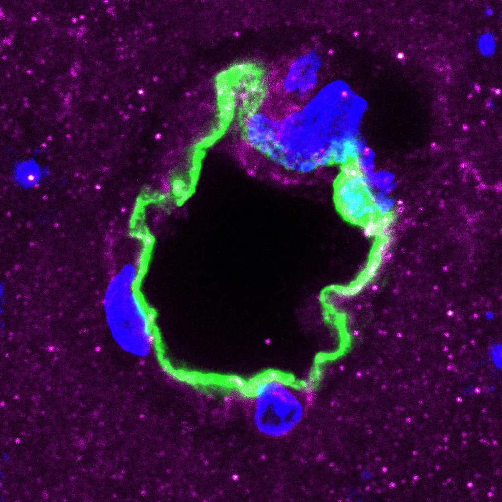
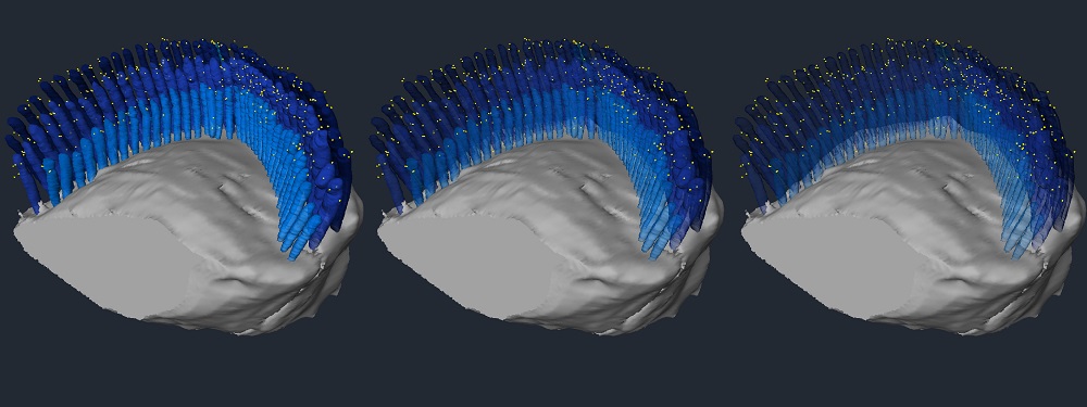
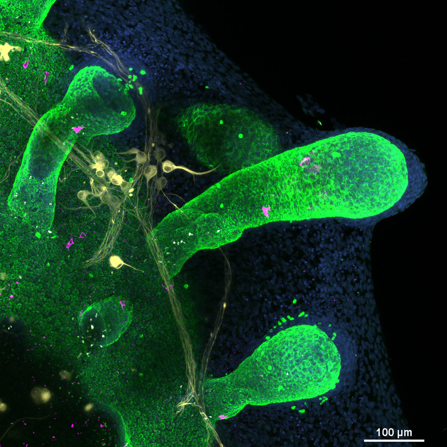
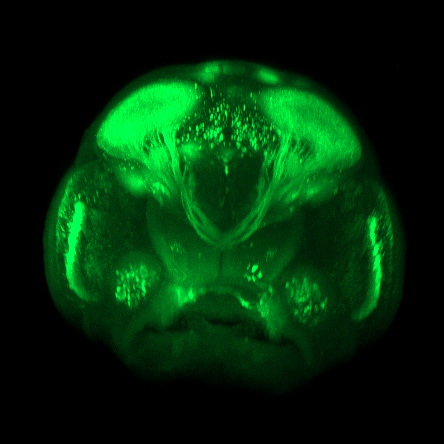
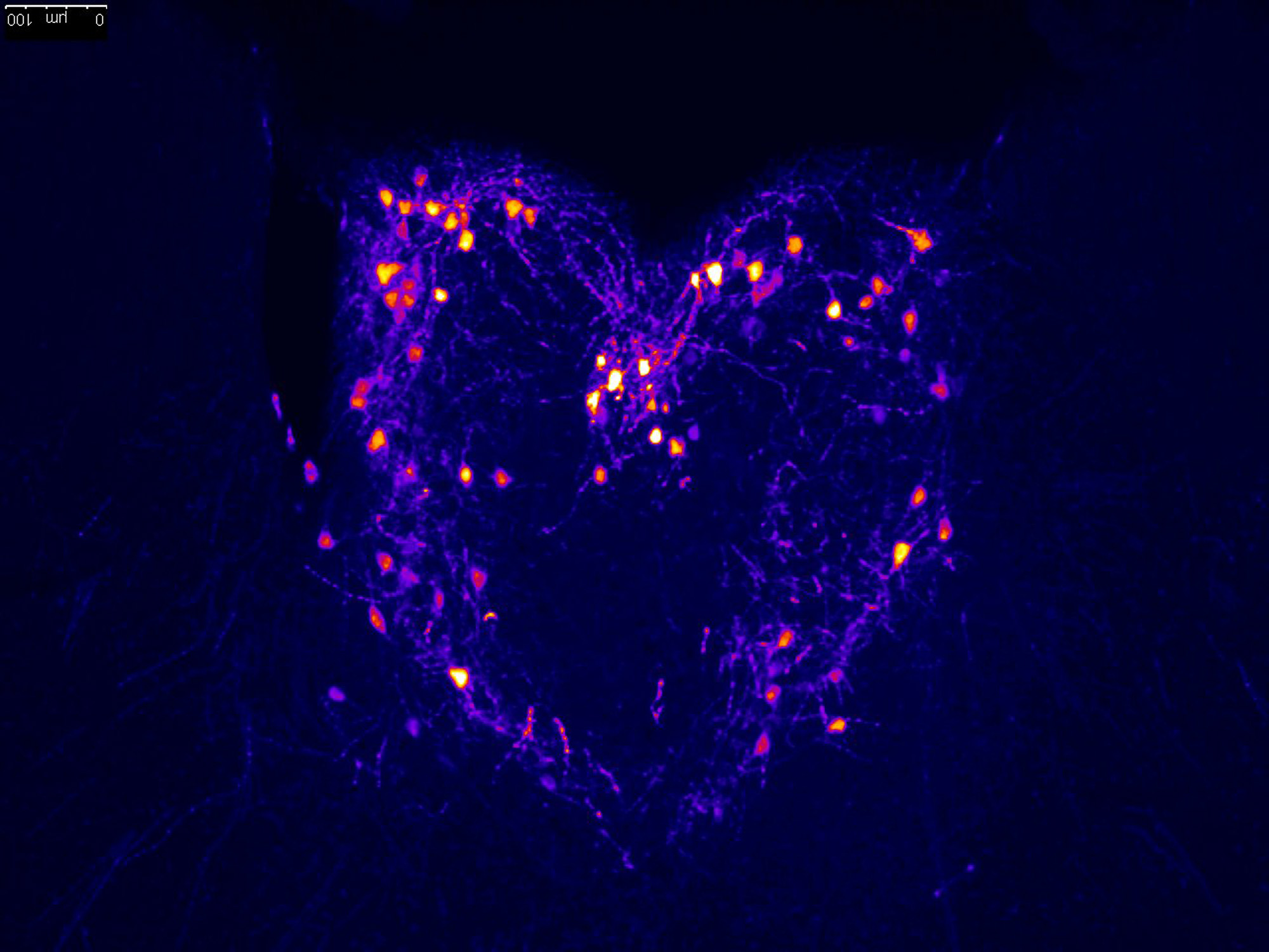
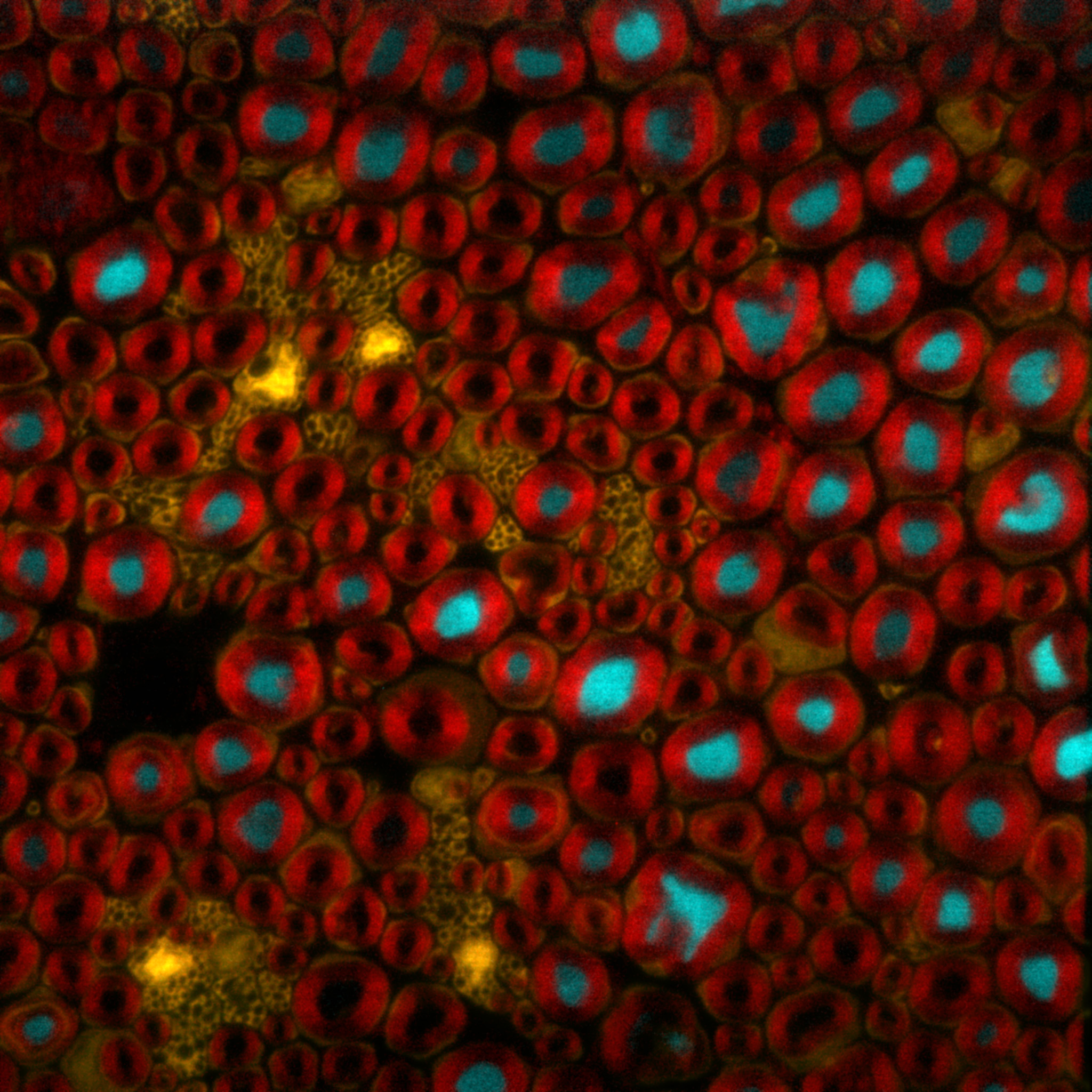
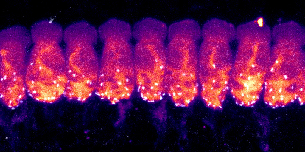
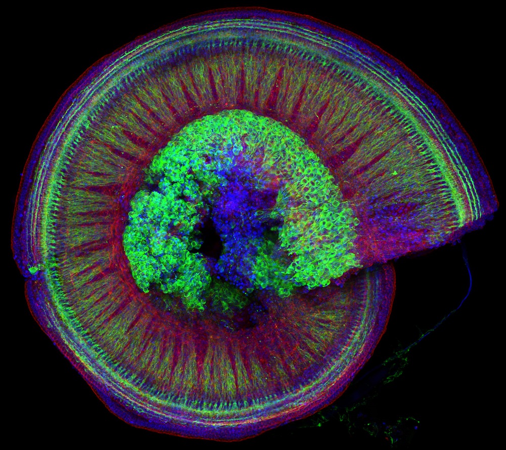
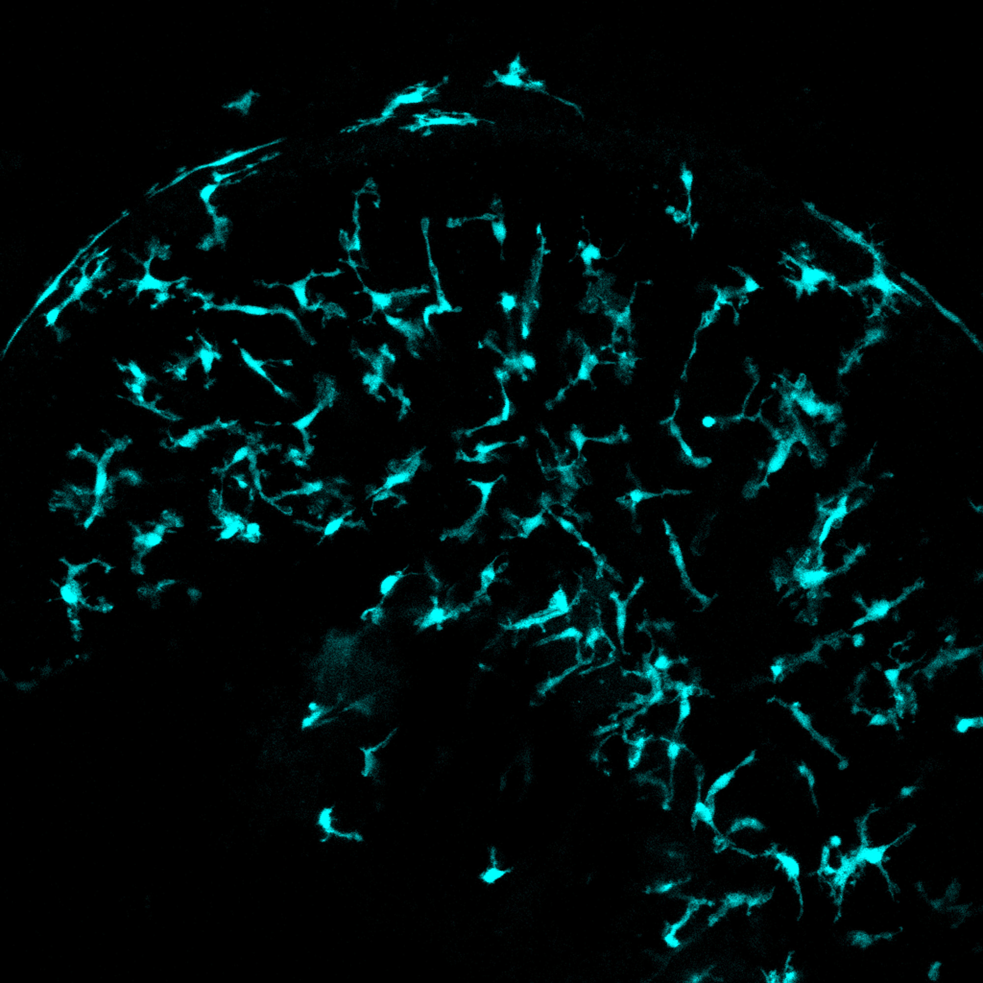
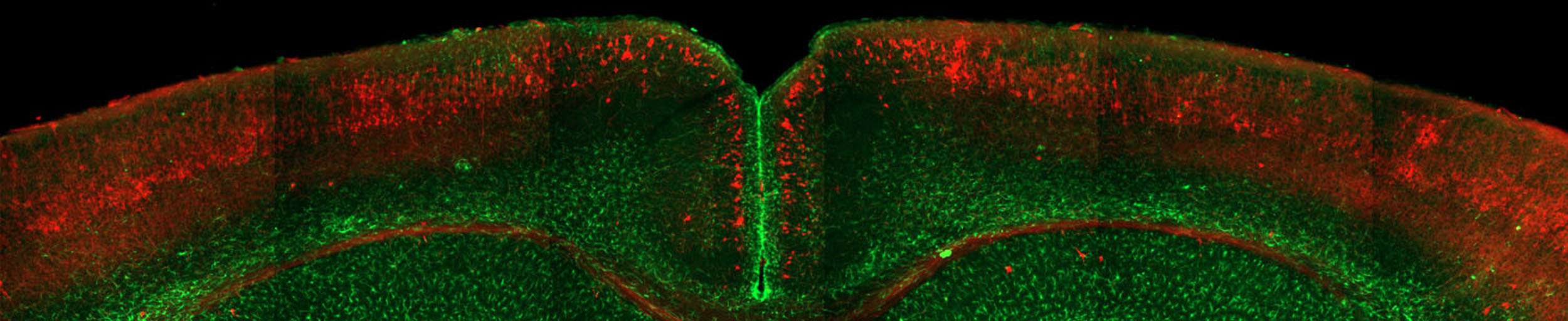
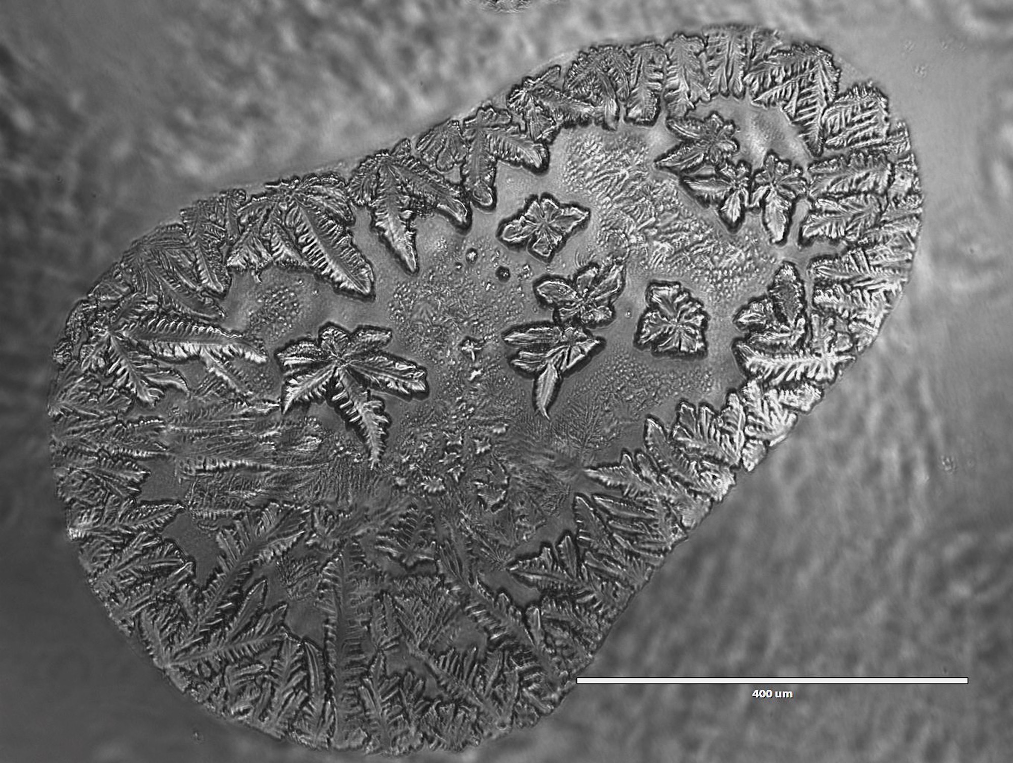
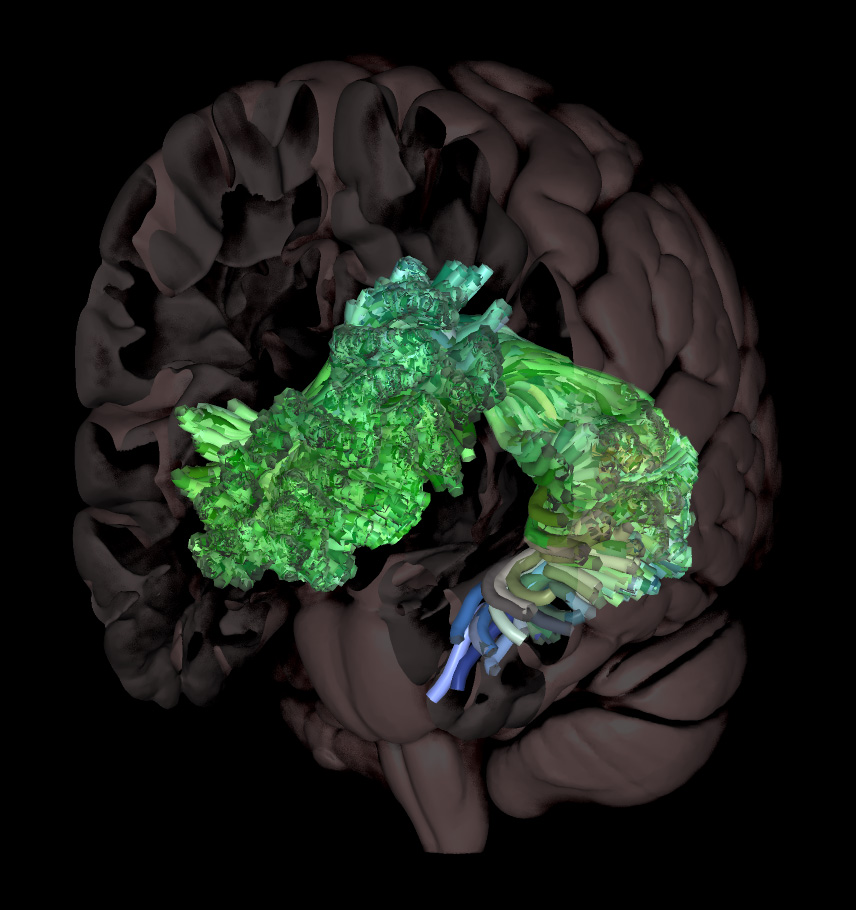
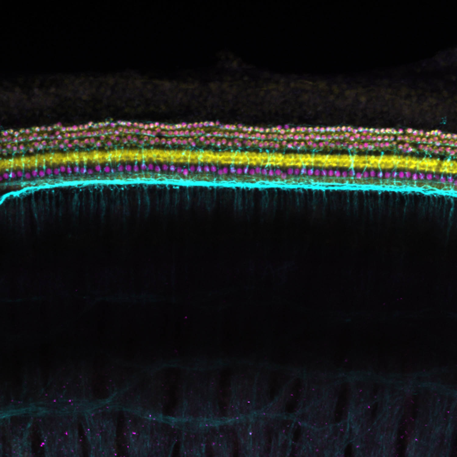
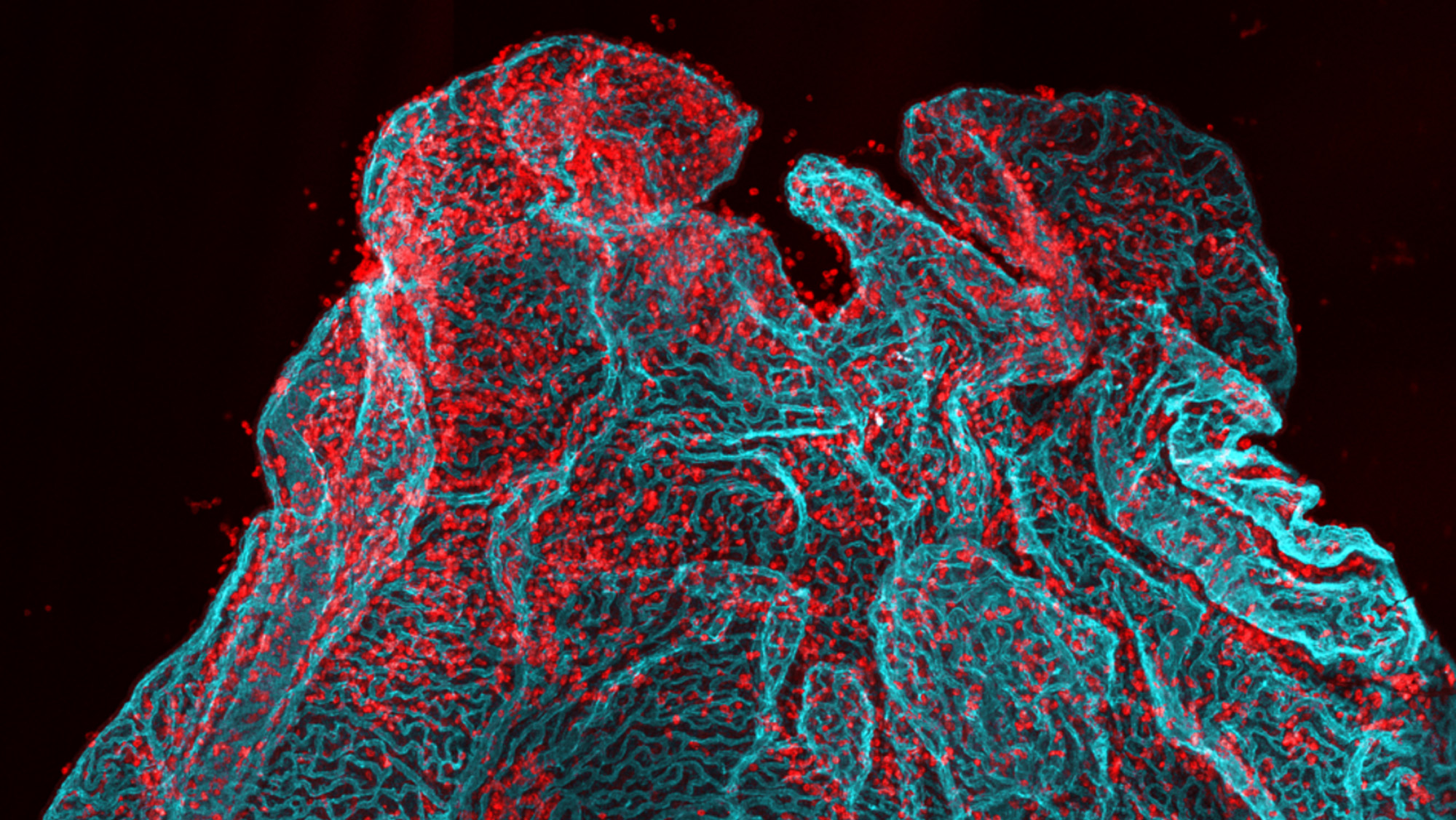
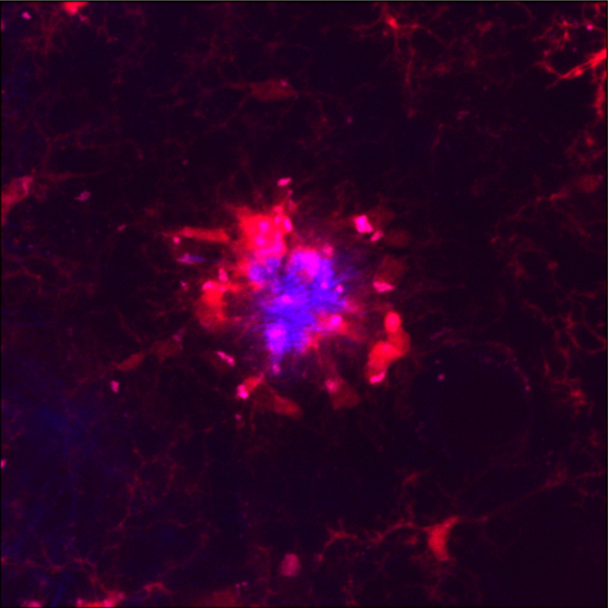
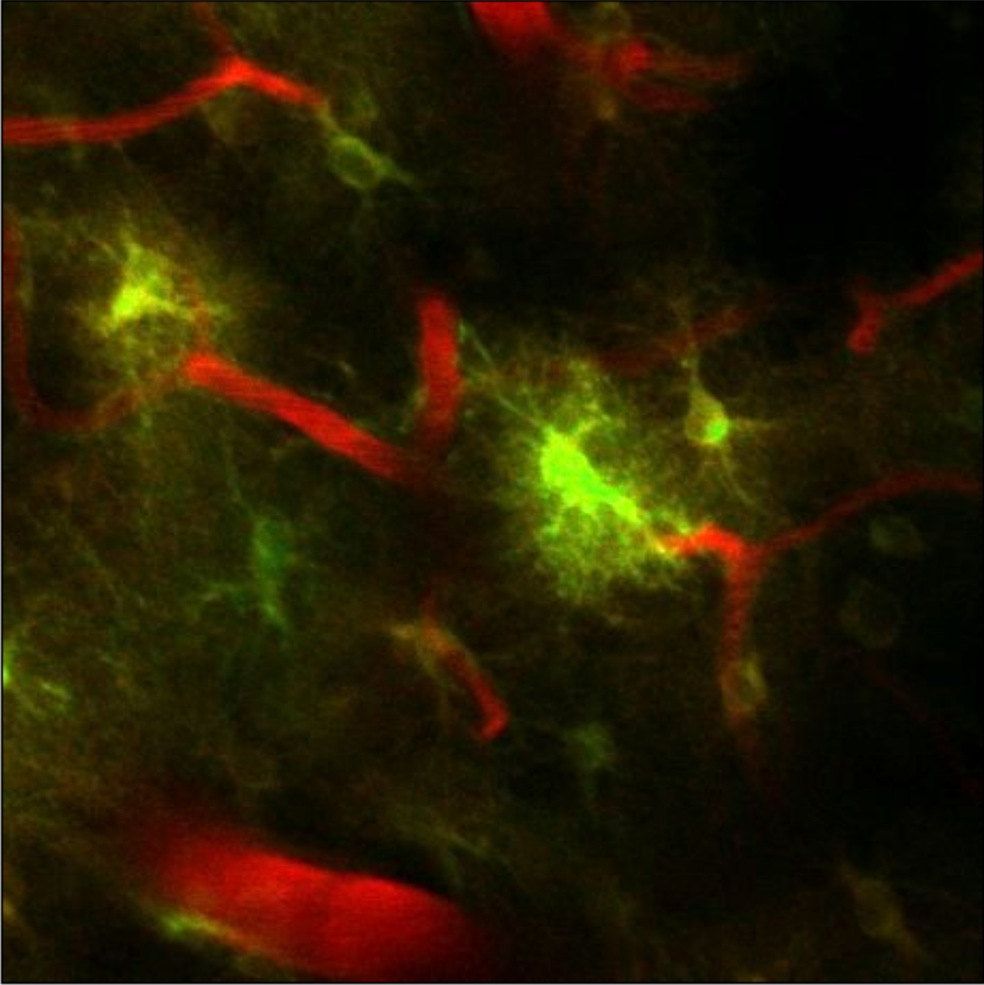
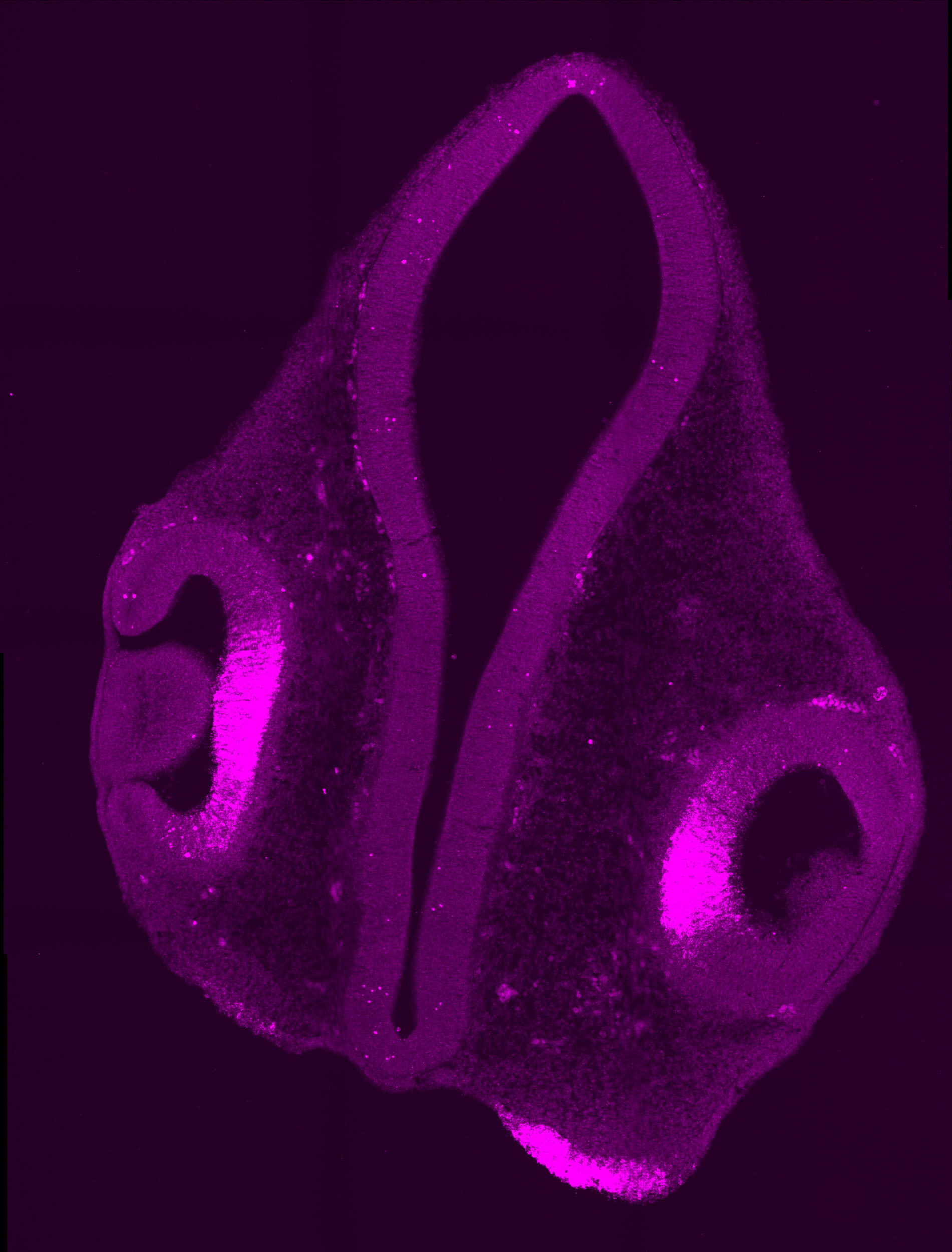
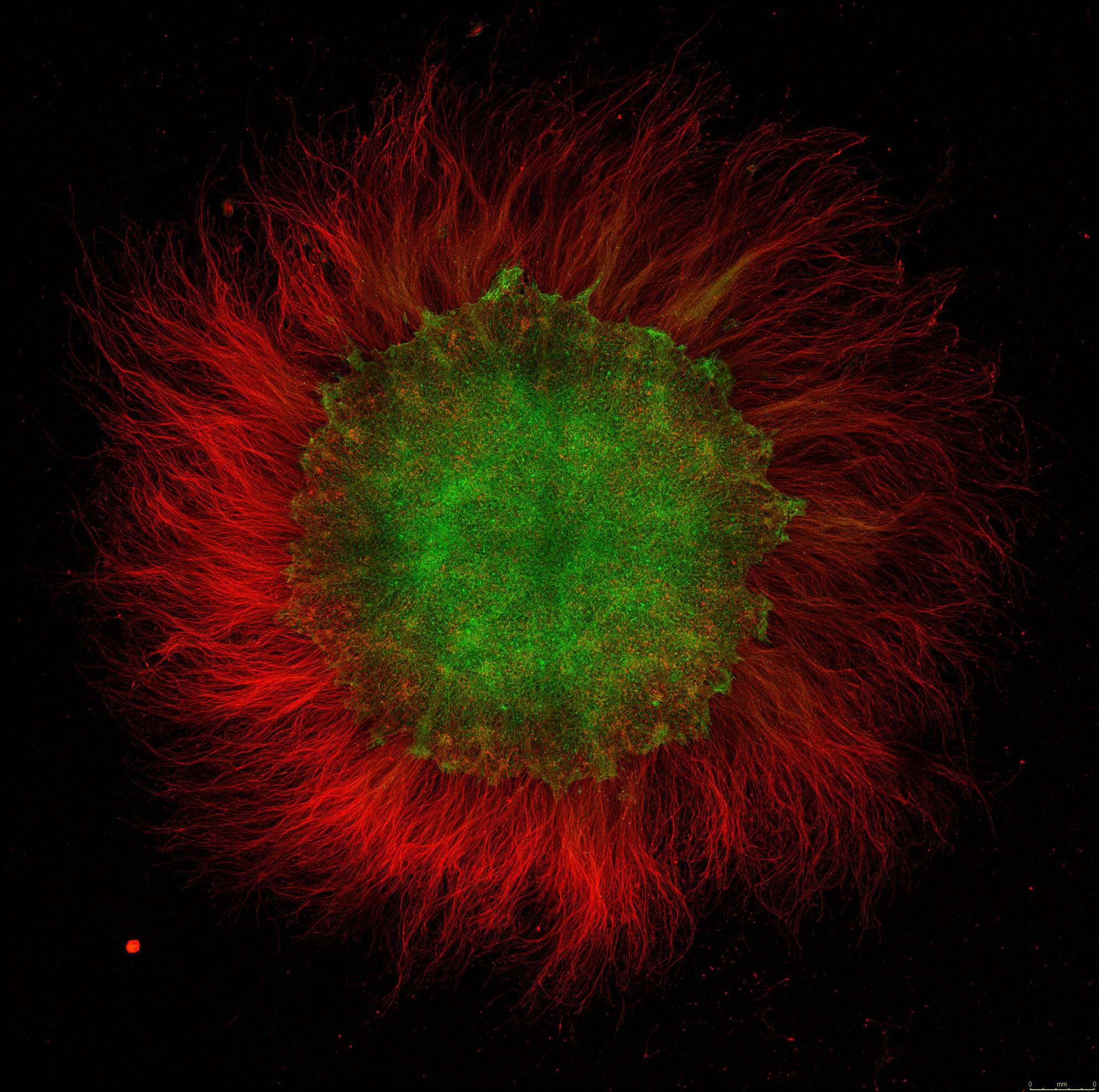
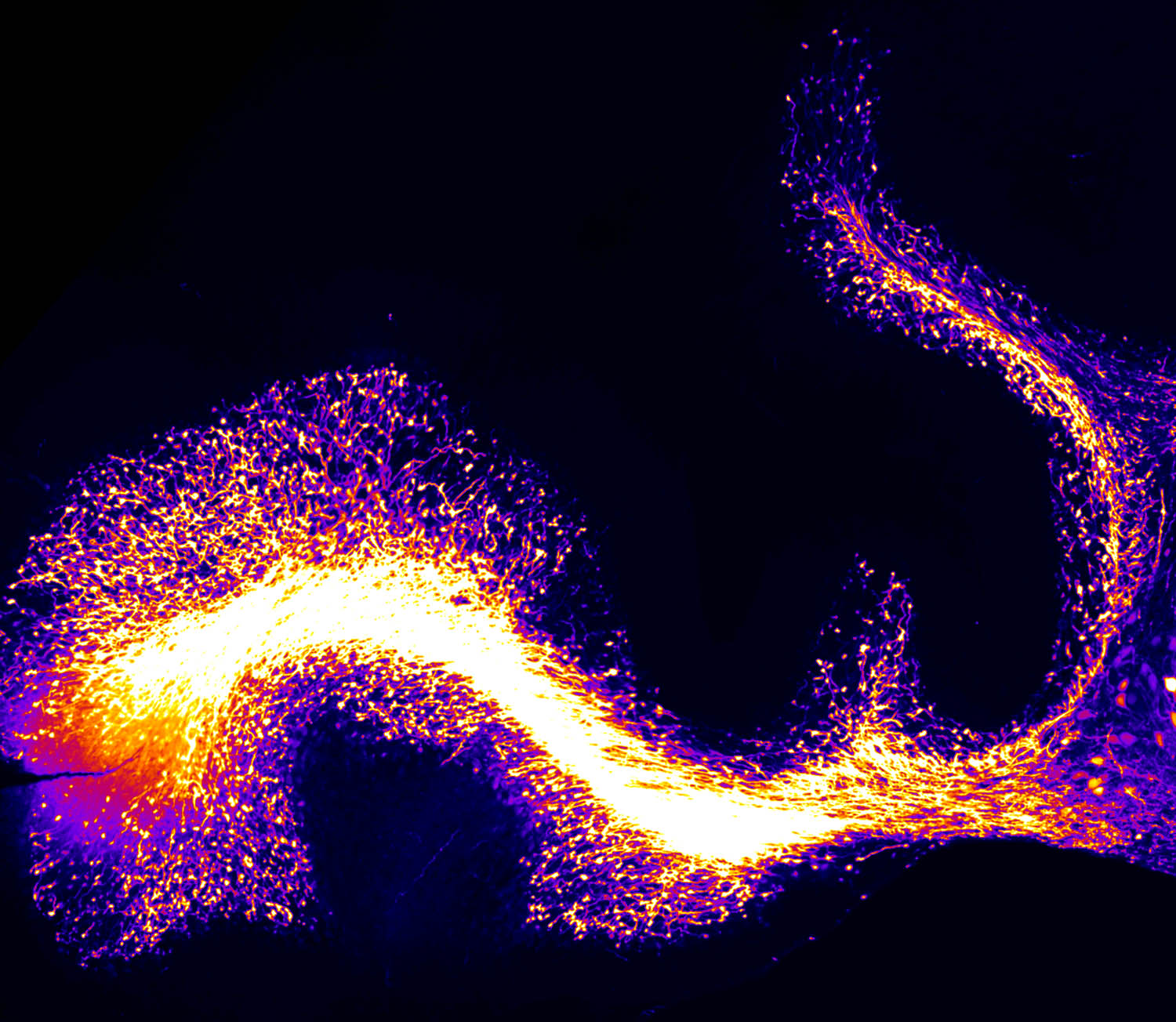
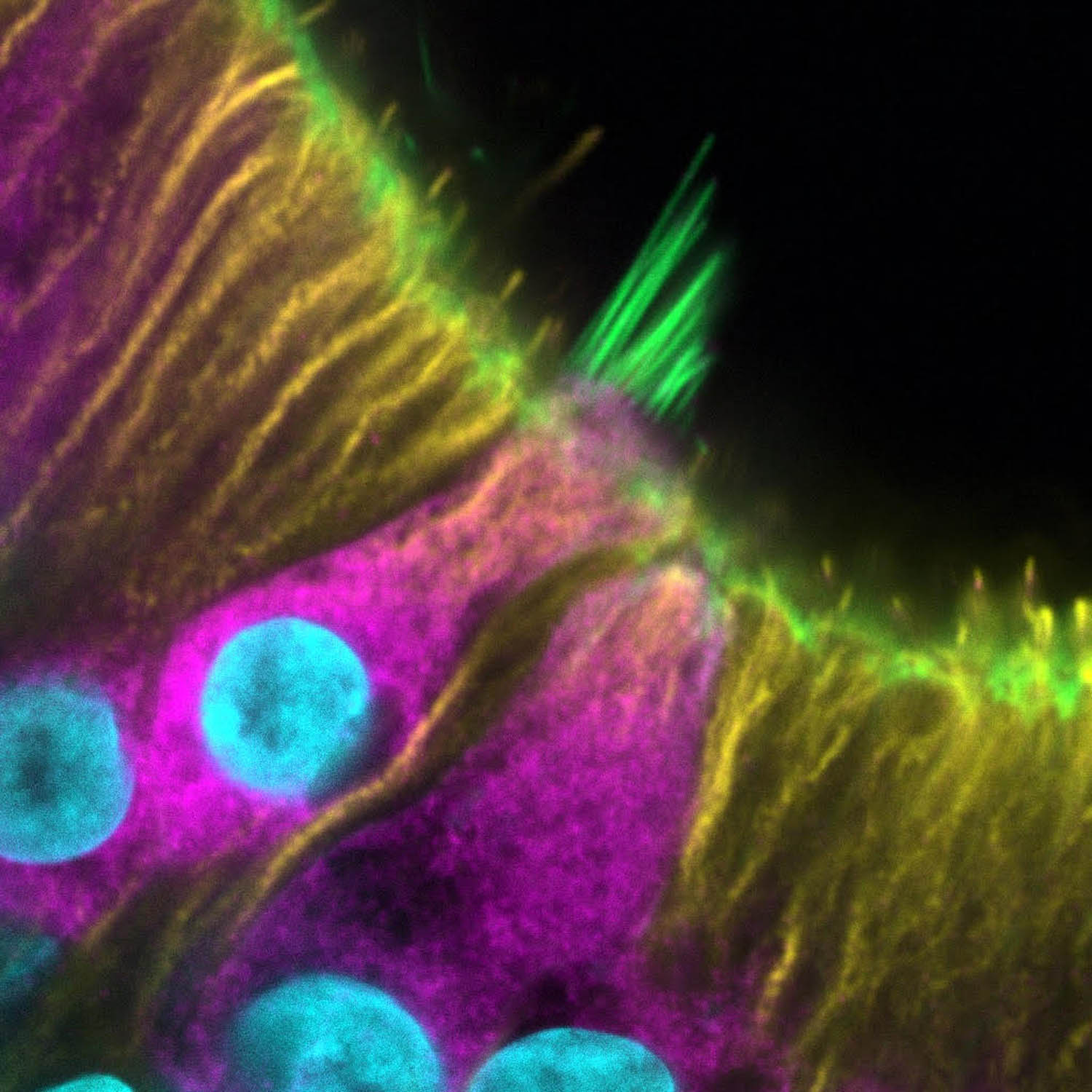
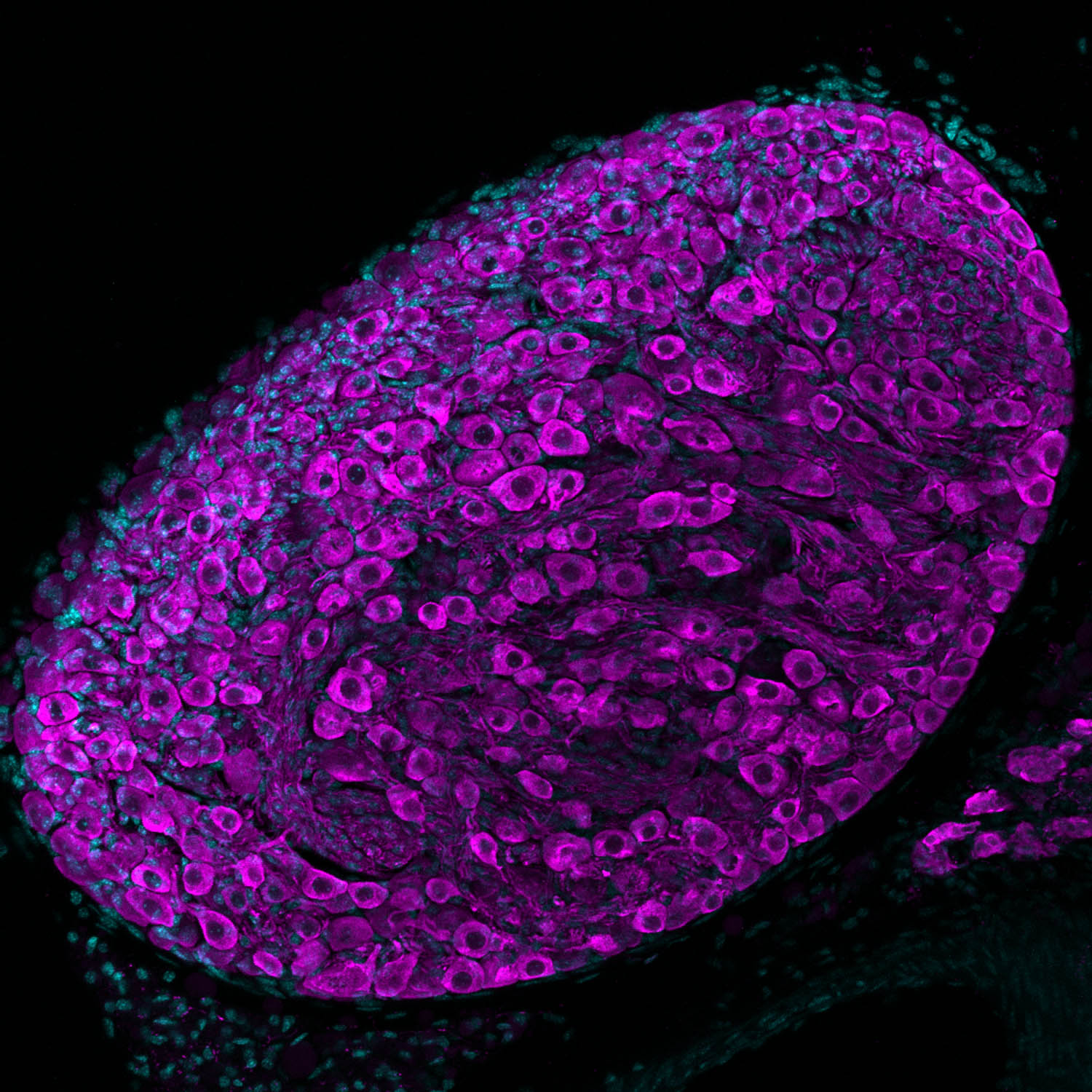
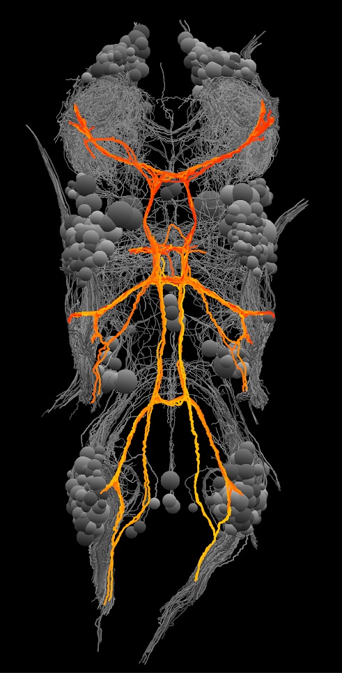
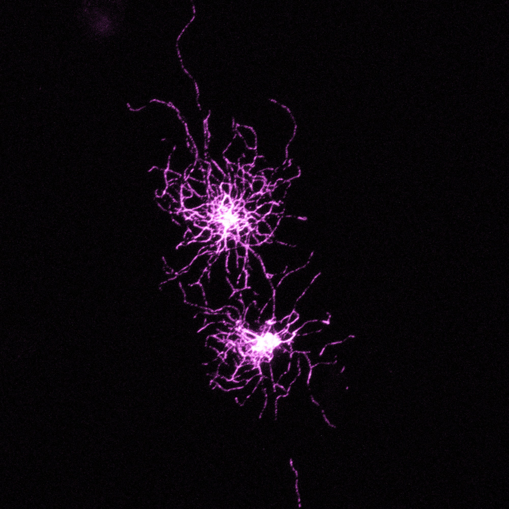
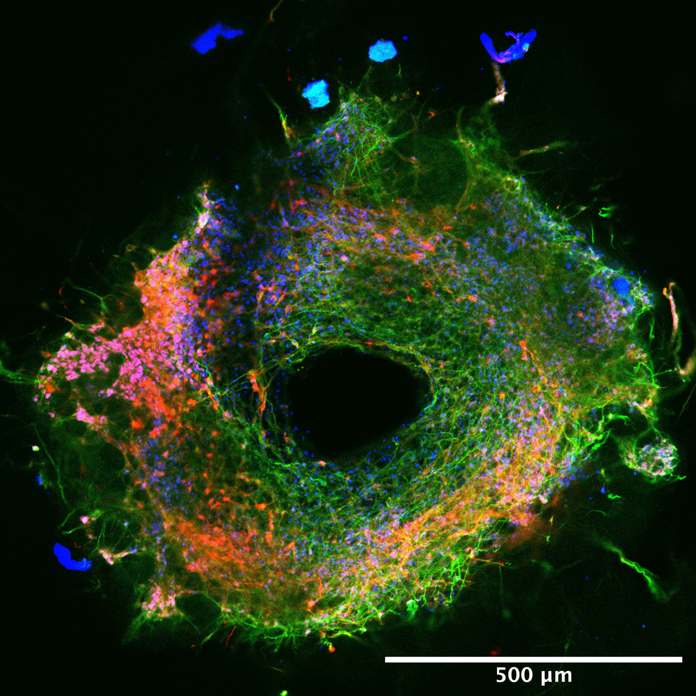
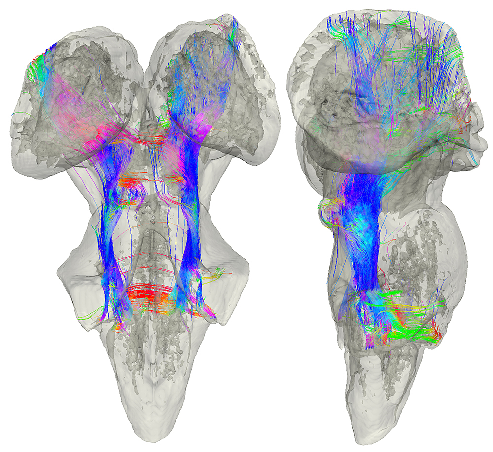
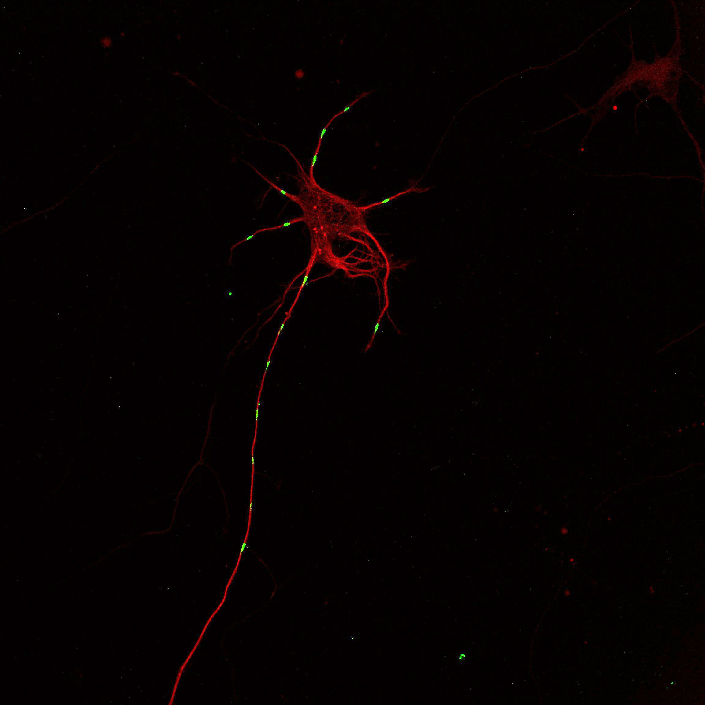
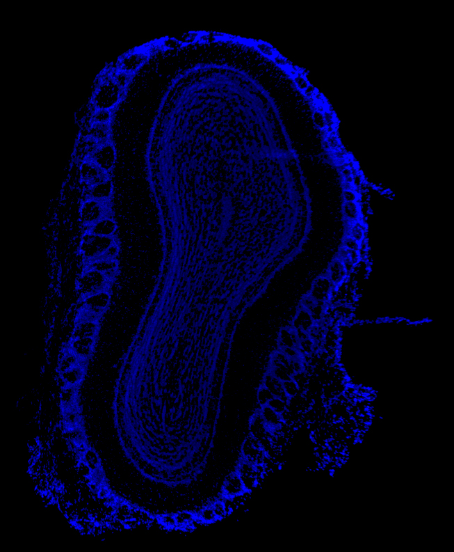
HBI Logo
  • About HBI
    • Our Mission
    • Contact Us
    • Mailing List
    • Support Us
  • People
    • Our Leadership
    • Our Staff
    • Faculty Directory
    • Community Directory
    • Humans of HBI
  • Science
    • Neuroscience at Harvard
    • Research Areas
    • HBI Grant Programs
    • HBI Community Groups
  • News
  • Events
    • HBI Events
    • Greater Boston Neuro Events Calendar
  • Resources
    • Mousekepedia
    • Beauty of the Brain
    • Photos
    • YouTube Channel
    • Core Facilities
    • Helpful Links
  • All images
  • 2025 Gallery
  • 2024 Gallery
  • 2023 Gallery
  • 2022 Gallery
  • 2021 Gallery
  • 2020 Gallery
  • 2019 Gallery
  • 2018 Gallery
This image displays parallel networks in parietal and temporal regions from 10 human subjects, highlighting the unique organization of association networks for working memory, remembering, language, and social inference. Distinct color patterns reveal how specialized brain networks vary across people, advancing our understanding of individual differences in brain organization. A Reconstructed Arcuate Fasciculus by Steven Meisler Into the Abyss of the Olfactory Landscape by Siddharth Jayakumar Boosting the Brain: The Choroid Plexus by Ryan Fame Pyramidals by Joseph Zak Integrating Granule Cells by Joseph Zak Scanning Electron Microscopy (SEM) image of the cochlea, the hearing organ, of a postnatal day 7 wild type mouse. This delicate structure is encased in the temporal bone of the skull and contains the specialized sensory cells (hair cells) and neurons (spiral ganglion neurons) that send sounds to the brain for downstream processing. Pericellular Basket Surrounds a Cell in the Lateral Septum by Rebecca Senft A human brain organoid (green/blue) surrounded by glioblastoma multiforme cell clusters (red). The cancer integrates into neural networks, showing how malignant cells infiltrate and rewire healthy brain tissue. This striking image captures the clash between organized neural architecture and the chaos of the most aggressive brain cancer. Rendered by Lynde Folsom All Orientations Welcome by Erin Diel Astrocytes in the mouse retina form a delicate, ramified web that weaves a complex cellular architecture. Human iPSC-derived Neuromesodermal Organoid by Delaney Wood Cochlear Immune Cells Phagocytose Bacteria by Katelyn Comeau Boulanger, A Fishy Nervous System by Morgan Phillips Confocal image reveals the complex beauty of human iPSC-derived cortical neurons cultured in a microfluidic chamber modelling Alzheimer’s disease. Fluorescently labelled neuronal RNA granules (red) travel along microtubule tracks (cyan), orchestrating the transport of translational machinery essential for localized protein synthesis, capturing both the precision and fragile elegance of neurons. Glia and Neurons in the Inner Ear by Isle Bastille Redefining Viral Infection by Tyler Krause Perineuronal nets glowing green through fluorescent microscopy, emerging from the extracellular matrix-the biological scaffold underlying neural circuitry. These structures embody the brain's central dilemma: stability to lock in essential connections, or plasticity to rewire and evolve, a choice that shapes every neural network. A Watchful Eye of NPY by Olga Alekseyenko Radial Journey by Katerine Morillo Unveiling Cerebellar Cytology with Dual Lenses by Xiaomeng Han The Brain in Time (Flower Representation) by William Orwig Front and back views of a human stem-cell-derived skin organoid. Fluorescent labeling reveals green hair follicles swirling like Medusa’s curls as the tissue self-organizes. Astrocyte Perched on a Capillary in Awake Mouse Brain by Patricia Kelly Synapses on Hair Cells by Isle Bastille TMS-EEG Source Space Colormaps in MMVT Outgrowth of the developing cranial motor nerves in an embryonic mouse at embryonic day 12.5. Cranial nerves are segmented and pseudocolored to allow for tracking of the nerves from their nuclei in the brain as they grow out towards their target muscles. Chick Retina-Bow by Masahito Yamagata This image shows neurons in the brain of a larval Japanese Rice Fish that transmit motor commands to the spinal cord. This was achieved by embedding a crystal of dye into the spinal cord and allowing the axons to carry dye to the brain. Color represents z-depth. Rising From Splashing Waves in a Thundery Night by Giacomo Maddaloni Spinal Cord Starry Night by Ilayda Alkislar This image shows a cleared zebrafish larva with all its nerves labeled using a fluorescent antibody.  A color scale indicates depth, transitioning from shallow nerves near the skin and fins to deeper ones innervating internal organs. Nemo’s Home: The Surface of a Human Inner Ear Organoid by Carl Nist-Lund Man-Made Brain by Annie Kathuria Ring in the Brain by Jaeeon Lee Medium Spiny Neurons in Ventral Striatum from a Behaving Mouse by Selina Qian On the left, a healthy brain organoid self-organizes into complex neuronal networks. On the right, the same system is invaded by glioblastoma multiforme (yellow), the deadliest brain cancer. Cancer cells integrate into neural circuits, exposing how malignancy hijacks human brain tissue and reshapes its delicate architecture. Nociceptors and Cancer Cells by Pavel Hanc Neuronal Plumage by Katherine Morillo C. elegans Nervous System by Rafael Jacobsen Sympathetic Ganglion Neurons by Katelyn C. Boulanger Neurons Within the Mouse Retina by Nicholas Hanovice Cerebellar Checkers by Ellen DeGennaro The Mind of a Worm Reconstructed by Electron Microscopy by Daniel Witvliet Hair Cells and Neurons of the Cochlea by Carl Nist-Lund Image 18 Flaming Cerebellar Cortex by Olga Alekseyenko This image shows two halves of a dog brain. In red we see a bundle of connections that process information about scent, mapped out by functional magnetic resonance imaging (diffusion MRI) studies. Scent processing takes up a lot of the dog brain! The Human Meissner Corpuscle by Ilayda Alkislar Neuronal Clusters by Evelyn Aviles Accidental Crystallization of Cell Media Solution on Culture Dishes Breast Cancer Cells in Co-Culture with Neurons by Rachel Davis On the Vine by Chelsey LeBlang The Murine Vomeronasal Organ and Immunity by Rodrigo Gonzalez A Sparkling Spot by Keunjung Heo The Site of Touch by Annie Handler The Intertwinement by Wardiya Afshar Saber and Federico Gasparoli Projection Neurons of the Fruit Fly Antennal Lobe by Matthew Churgin Distribution of N- and O-GalNAc Glycans in the Cerebellum by Maxence Noel This image shows Pacinian corpuscles in the mouse hindlimb, each innervated by an axon (magenta), with supportive cells forming an inner (green) and outer corpuscle (red). The outer corpuscles touch, likely to assist signal transduction to the central nervous system. Pluripotent Stem Cell Derived Neural Tube-Like Organoids 1 by Dosh Whye Brain Tumor of a Fruit Fly by Torrey Mandigo Retinofugal Projections into the Optic Tectum of Larval Zebrafish by Clemens Riegler Plaque Attack by Aryan Rajput Sole Projector by Leannah Newman. Actin Network Architecture of a Motor Neuron by Iván Coto Hernández Cervical- and Lumbar-Projecting Descending Projection Neurons in the Red Nucleus by Carla Carol Winter Spinal projection neurons of larval Japanese Rice Fish (left) and Zebrafish (right) labeled with z-depth color code. These fish diverged 300 million years ago, yet the basic neuronal architecture for motor command transmission is highly conserved. Astrocytic Hug by Aryan Rajput Clock Neuron Subpopulations in Drosophila melanogaster by Bryan Song A Cloudy Memory by Leana Radzik Network at the Fingertips by Sara Hakim and Yu-Ting Cheng The mammalian cochlea is a spiral-shaped organ that mediates the sense of hearing with astounding precision. Here, auditory neurons are shown with their cell bodies in magenta, extending cyan axons to innervate sound-sensitive cells around the spiraling border of the tissue, also in magenta. Thoughts and Feelings by Paul Hatini Podoplanin in the DRG by Pavel Hanc The Olfactory Mantle by David Brann This image shows a cross section of an early chicken head at a stage when the eye is just beginning to form. The bright signal marks FGF8 at the center of the developing optic cup. FGF8 is a growth factor (?) that plays a critical role in organizing the future high-acuity region of the retina—the area responsible for sharp, detailed vision in adults. Even at this very early stage, key molecular cues are already shaping where specialized visual functions will emerge. The image highlights how early developmental signals help lay the blueprint for the development of key aspects of visual function long before the eye can detect light. Microglia (green) remodel synaptic connections, while inhibitory neurons (red) maintain network balance. This human stem cell–derived co-culture is used to investigate how microglia refine inhibitory synapses to tune neural communication. Ear to Hear: Spiral Ganglion Neurons of the Cochlea by Katelyn C. Boulanger Spiny Night: A Neuron Illuminated by Maryse Thomas Astrocyte Waltz by Molly Healey This whole-mount projection of a chemically cleared mouse spinal column visualizes the spatial relationship between skeletal and vascular systems. Mineralized bone forms a continuous axial chain (red), while CD31-immunolabeled endothelial networks (blue) course along the vertebral bodies. The resulting tapering profile and interlaced vascular contrast incidentally evoke the appearance of a dragon's tail. Unipolar Brush Cells Contacted by Purkinje Cell Feedback by Chong Guo Distribution of GJB2 in a Nonhuman Primate Cochlea by Maryna Ivanchenko From Human Adult Fibroblast to CA3 Neuron by Haley Zanga Guardians of the Brain Barrier by Huixin Xu Local interneurons in the drosophila antennal lobe by Isabel D’Alessandro Wheel in Chick Retina by Masahito Yamagata Ferret Phases by Ellen DeGennaro Serotonergic Modulation of Sensory Information by Olga Alekseyenko Neuronal Network Cultured on an Integrated Circuit Chip by Jeffrey Abbott and Tianyang Ye The Circle of Willis by Brian Chow In the skin, sympathetic nerves, arrector pili muscles, and hair follicles form a tri-lineage unit that produces goosebumps and regulates hair follicle stem cell activity in response to cold exposure. Scanning Electron Microscopy (SEM) image of two turns of the cochlea, the hearing organ, of a postnatal day 7 wild type mouse. On each turn, four rows of specialized sensory hair cells can be seen. Hair cells are responsive to frequencies of the sound spectrum in a structured tonotopic organization from base (high frequencies) to apex (low frequencies). The Brain in the Gut by Valentina Lagomarsino Wholemount of the Mouse Cochlea by Isle Bastille Human Pluripotent Stem Cell-Derived Cortical Organoids by Dosh Whye In the inner ear, three populations of feedback neurons intermingle with the fibers of auditory neurons to change how our ears respond to stimuli. Their axons display a range of appearances but still are very organized, bringing to mind strings of holiday lights of different colors all arranged with care. Let's Play Catch! by Methasit Jaisa-Aad Microglia (red) populate the proximal optic nerves of a mouse with optic pathway glioma. The putative tumor-bearing region (right) shows disrupted cellular organization, where irregularly positioned oligodendrocytes (pink) trace the altered architecture of the optic nerve. Multicolor Imaging of Peripheral Nerve by Iván Coto Hernández Organized Mess by Stephanie Mauriac Mouse Dorsal Root Ganglia Coronal Section of the Dorsal Raphe Nucleus of a Mouse Midbrain by Olga Alekseyenko Staying in Touch by Urs Langen Vasculature in the Developing Cochlea by Katelyn Comeau Steering Control by Emily Kellogg Giving Feedback by Austen Sitko Intersubject Synchronization of Brain Activity During Viewing of Film Clips by Xuehu Wei The image depicts EB3–GFP–labeled microtubule plus-end tracking proteins in a cultured mouse hippocampal neuron. Astro Heart by Elisabeth Lawton Kiss in a Starry Night: Overcoming Developmental Constraints by Giacomo Maddaloni The Brain in Time (Snake Representation) by William Orwig Lateral Olfactory Tract by David Brann Olfactory Bulb Glomeruli by Joseph Zak Cortical Sparks by Lariza Rento A motoneuron-containing midbrain organoid derived from human induced pluripotent stem cells (reprogrammed from skin tissue), cultured in vitro for 55 days. The magenta staining (Islet1) reveals the organized nuclei of motoneurons, the blue staining (ChAT) marks cholinergic neurons, and the green staining (MAP2) specifically labels neuronal microtubules. Pluripotent Stem Cell Derived Neural Tube-Like Organoids 2 by Dosh Whye A confocal microscopy image showing human neurons (magenta) forming connections with diffuse midline glioma cells (green), a rare childhood brain cancer. The red and white dots mark synapses — tiny contact points where the cells communicate. These signals show how tumor cells interact with and respond to nearby neurons. Blood Vessels in the Developing Retina by Swathi Ayloo Gray Matter Galaxy by Ayush Noori and Clara Muñoz-Castro Hair Follicle With Touch Receptor Cells by Ahn Phuong Le A map of a growing neuron's molecular highways. Microtubules (red) make up the neuron's structural support and transportation routes. Patches of microtubules (green) have been converted to a different color to track their movements. A coronal section of an adult mouse brain. Green staining marks astrocytes expressing glial fibrillary acidic protein (GFAP), while red indicates the expression of a cell membrane-targeted protein in layer 2/3 cortical neurons. Reaching Out by Austen Sitko Chain of Olfactory Glomeruli by Nao Horio Fish is Watching You 2 by Yasuko Isoe Lightsheet Illuminating Our Knowledge of the Brain by Andrew Silberfeld Human iPSC-derived Neuromesodermal Organoid by Delaney Wood This image was captured as part of our research into understanding nerve–stem cell interactions in the skin. Shown here is the intricate network of sensory neurons (blue) that innervate the skin epidermis and hair follicles (white), enabling us to perceive touch, temperature, and pain—crucial functions for adapting to a constantly changing environment. Cluster of E1 Neuronal Population in a Virgin Male Mouse by Samantha Finkbeiner A Reconstructed Corticospinal Tract by Steven Meisler A coronal slice of an adult mouse brain showing antibody staining for glial fibrillary acidic protein (GFAP), which labels reactive astrocytes in the adult brain. Anti-Amyloid Antibody Binding to Vascular Amyloid with Microglia Recruitment by Praveen Bathini. mEMbrain by Elisa Pavarino Let There Be Sound by Volkan Ergin Human Brain Spatial Transcriptome by Rongxin Fang Auditory Rainbow by Stephanie Mauria Developing Zebrafish by Barbara Robens Festive Fluorescence by Marlene Scheffold This confocal image captures the balance that keeps the brain in tune. In the mouse frontal cortex, a delicate network of dendrites (blue) weaves together with inhibitory synapses marked by VGAT (green) and gephyrin (red). These vibrant threads of inhibition reveal the subtle architecture that tempers and fine tunes neural activity to maintain balance within the brain’s circuitry. Merry Christmas from iPSCs by Wardiya Afshar Saber and Federico Gasparoli Star-Crossed by Elisabeth Lawton Stem Cell-Derived Human Hair Cell of the Inner Ear by Carl Nist-Lund Developing Motor Neurons and Muscles in E11.5 Mouse by Jess Bell & Mary Whitman A straight-on view toward the eye of an embryonic mouse showing the extraocular muscles (magenta) as the oculomotor (cyan) and trochlear (green) nerves reach the area and connect to their target muscles. Cells and Vessels by Mostafizur Rahman E11.5 Orbit Showcasing Developing Extraocular Muscles and Cranial Nerves by Jess Bell & Mary Whitman Amyloid Coral Reef by Celia Blanco. Developing Zebrafish by Barbara Robens Brain Star Dome by Ayush Noori and Clara Muñoz-Castro The Hypothalamus Revealed: Fluorescent Luminance Meets Ultra-Structural Detail by Xiaomeng Han. Six-Color Immunolabeling of Mouse Cerebellar Cortex by Xiaomeng Han Innervation of a Hair Follicle by Touch-Sensitive Neurons by Katelyn Comeau Cervical- and Lumbar-Projecting Corticospinal Neurons Carla Carol Winter Genetically Labeled Tapestry: Coloring the Brain Net of the Gut by Anoohya Muppirala his image shows Pacinian corpuscles in the mouse hindlimb, located along the fibula, which each have an inner corpuscle composed of terminal Schwann cells (green), innervated by a single peripheral axon (magenta). Sketching the Brain by Minsu Kim Mapping of Superficial White Matter Connections: by Fan zhang Love From Your Neurons by Encarnacion Torres Losing Stability in Bipolar Disorder by Katharina Meyer This image captures the glowing mitochondria inside a rat neuron, shining like tiny embers. These bright fragments are the cell’s “power plants,” traveling along its branches to deliver the energy that makes every thought, memory, and movement possible. Man-Made Brain by Annie Kathuria Spiral Ganglion Neuron Diversity by Isle Bastille Rare Perspectives on Rare Diseases by Marlene Scheffold Proximity of BMAL1 and CaMKIIα at Synapses in a Primary Neuron by Ilaria Barone In the inner ear, immune cells make very close contact with pericytes, cells that wrap around blood vessels so small that red blood cells have to pass though in single file. These vessels weave through neuron fibers as they reach out to hair cells that sense vibration (top). Diffusion MRI Tractography of the Human Subcortical Auditory Pathway by Kevin Sitek Glowing mitochondria dot the long branches of neurons, highlighting the cell’s internal transport system. These mobile energy producers travel up and down the neuron to keep it functioning, powering the signals that allow brain cells to communicate over remarkable distances. Accidental Crystallization of Cell Media Solution on Culture Dishes by Farimah Mapar Caught in the Net by Carolyn Johnson Trafficking of RNA Molecules to the Growth Cone by Kadir Ozkan Breaching of the Choroid Plexus Barrier by Huixin Xu In white are mouse retinal ganglion cells, the output neurons of the eye that communicate with the brain. In red are retinal bipolar cells, interneurons which relay information from photoreceptors to retinal ganglion cells. Stairway to Heaven: The Bundle of a Human Stem-Cell Derived Hair Cell by Carl Nist-Lund Connect to Hear: Afferent and Efferent Neurons in the Ear by Yi-Chia Huang Red Blood Cells in Retina Vasculature by Swathi Ayloo This image depicts a cross-section of the mouse nose after treatment with a drug that ablates the olfactory tissue, stained for a sensory neuron marker gene (in magenta) and specific subtypes of olfactory receptors (in yellow ). The expression of these genes and their spatial patterns indicates proper regeneration. Miami on the brain by Natasha M. O'Brown The dura is the outermost layer of the meninges, a membrane that covers and protects the brain. This image shows a small molecular tracer NHS-biotin (gray) leaking out of blood vessels (green) in the dura. All cell nuclei are stained in blue. Magenta labels a histone modification that is associated with transcriptionally inactive DNA. Skin Organoid Generated From iPSC with Innervated Neurons by Ahn Phuong Le Neuronal Stem Cells Differentiating Into Neurons by Katharina Meyer Mapping of Whole Brain White Matter Connections by Fan Zhang Dancing Hairs by Cristobal Von Muhlenbrock Rodriguez Neuron-Painted Portraits by Will Xiao Enteric neurons reside within the gut, so numerous that they have been referred to as The Starburst Amacrine Cells of the Retina by Evelyn Aviles Neurons and Sensory Cells of the Mouse Cochlea by Katelyn Comeau AH5 Coronal Slice 1127 by Javier Masis Tangled Tubules by Mara Casebeer Spinal Motor Neurons by Spencer Price Fish is Watching You 1 by Yasuko Isoe Superficial White Matter Connections to Broca’s Language Area by Shiva Hassanzadeh-Behbahni A 3D Reconstruction of an Anti-PKHD1L1 Immunogold Labeled OHC Stereocilia Bundle A Touch of Fire by Lucy Gordon Twinkle Stars in Alzheimer’s In-a-Dish by Sang Su Kwak Synaptic Talk Inside the Brain by Nannan Guo Immune Cell Surveillance by Katelyn Comeau Boulanger Cortical neurons derived from human induced pluripotent stem cells (reprogrammed from skin tissue), grown for 14 days in a dish. The rose-like structures are called neuronal rosettes. The blue staining marks all cell nuclei, the red staining marks microtubules, and the green staining reveals expression of one transcription factor specific to cortical neural progenitor cells (FOXG1). Neurofilaments by Evelyn Aviles Symmetric Division of a Neural Stem Cell by Haley Zanga Brain Waves by Lariza Rento Immune cells (green) are shown interacting with blood vessels (magenta) within the dural layer of the meninges, a membranous structure that covers and protects the brain. This close interaction between immune cells and the vasculature highlights the role of the dura in immune surveillance and neuroimmune communication. All nuclei are shown in blue. Neurons that Sense Blood Pressure in the Aortic Arch by Soohong Min Human Retinal Organoids to Study the Underlying Causes of Spinocerebellar Ataxia 7 by Patrick Ovando-Roche Aurora Hippocampus by Riley Magane and Meng Chen Krause Corpuscles in the Mouse Clitoris by Lijun Qi The skin of a mouse ear by Brian W. Chow Astrocyte-Blood Vessel Interaction by Urs Langen Before thought begins, the fetal brain performs its first selection, discarding fractured genomes to preserve the integrity of those destined to shape the self. Blue DNA escapes through the orange nuclear envelope of a developing neuron, forming micronuclei, tiny archives of genetic failure marked by green rupture signals. vDeltaA Neurons in the Fly Brain by Emily Kellog Vessel near ENS by Valentina Lagomarsino Anti-Amyloid Antibody Binding to Vascular Amyloid with Microglia Recruitment by Praveen Bathini A patient induced pluripotent stem cell-derived neuroepithelial organoid. Neuroepithelial stem cells self-organize in a columnar-like fashion around a central cavity (lumen). This process mimics the early spatial organization of the developing neural tube. Live 3D cell imaging highlights Plasma membranes (Red) and Nuclei (Blue). 3D sensory neuron organoid with cell bodies clustering in the middle and radiating out axonal projections, stained for the building block, tubulin, with a live dye - SIR tubulin Bed of Roses by Katharina Meyer Art Generated by Neurons (no. 6) by Will Xiao A Portal by Lucy Gordon In Vivo Organ of Corti by Nam Hyun Cho Glial Diversity in the Olfactory Bulb by Anoohya Muppirala Love of Hearing by Jin Jiahe Multicolor FlpOut Clones of Drosophila Ellipsoid Body Ring Neurons by Isabel D’Alessandro Neural Hyperspace by Jenny Yau Human hippocampus, the brain region where memories are consolidated. Astrocytes were stained with the cytosolic marker ALDH1L1 (green) and the endfeet marker AQP4 (red). All nuclei were stained with DAPI (blue). Blood vessels are an essential component of all organ systems, as they supply cells with oxygen and nutrients necessary to develop and function. Here, the developing vasculature (in red) is observed invading the embryonic cochlea (our hearing organ, outlined in dark blue), where it supports developing auditory neurons (in cyan). A cross-section of the mouse cochlea, the organ that detects auditory information. Sound-receiving inner hair cells and hearing-related neurons are shown in cyan, with brightness differences indicating varying expression levels. Pain-sensing neurons are labeled in magenta, revealing the mixture of sensory cells within the cochlea. Expression of PCDH15 in a Nonhuman Primate Photoreceptor by Maryna Ivanchenko Motor Neurons Innervating Eye Muscles by Sampath Vemula Astrocyte-blood vessel interaction by Urs Langen Confocal image reveals the complex beauty of human iPSC-derived cortical neurons cultured in a microfluidic chamber modelling Alzheimer’s disease. Fluorescently labelled neuronal RNA granules (red) travel along microtubule tracks (cyan), orchestrating the transport of translational machinery essential for localized protein synthesis, capturing both the precision and fragile elegance of neurons. Birth of New Neurons in the Adult Mouse Brain by Brittany Mayweather An isolated microvessel (a tiny blood vessel) from donated human brain, highlighted in light blue (using a cell membrane stain, Concanavalin-A). In yellow, you can see the Slices Under Stress: Neuronal Activation in the Mouse Brain by PK Vincze Glomeruli of the Fruit Fly Antenna Lobe by Matthew Churgin Sensory Tulips Jin Jiahe An isolated microvessel (a tiny blood vessel) from donated human brain, highlighted in light blue (using a cell membrane stain, Concanavalin-A). An astrocyte is hugging this microvessel, highlighted in yellow (stained for GFAP, an astrocyte marker). This image shows the intimate interactions between two different cell types in the brain. Unique Distribution of N-Glycans in the Brain by Maxence Noel Human iPSC-Derived Spinal Spheroid by Keunjung Heo This confocal image captures neurons in the mouse dentate gyrus, a region of the mouse hippocampus where new neurons emerge and memories form. Neuronal dendrites and cell bodies (MAP2, magenta), neuronal nuclei (NeuN, green), and all cell nuclei (blue), are shown composing a fluorescent landscape of connectivity and growth. Brain Teardrops by Methasit Jaisa-Aad Migrating Neurons in Embryonic Mouse Cortex by Kadir Ozkan A map of a growing neuron's molecular highways. Microtubules (red) make up the neuron's structural support and transportation routes. Patches of microtubules (green) have been converted to a different color to track their movements. Non-Neuronal Network in the Developing Cochlea by Olubusola Olukoya. The Inner Ear Rainbow by Cristobal Von Muhlenbrock Rodriguez When the Heart Finally Catches Up to the Brain by Encarnacion Torres Jimenez Sensory and Motor Neurons in the Drosophila Nerve Cord by Jasper Phelps Different Types of Cells in the Cerebellar Cortex by Xiaomeng Han Nose to Brain by David Brann Glia and Neurons in the Inner Ear by Isle Bastille Unipolar Brush Cells Contacted by Purkinje Cell Feedback by Chong Guo Touch-Sensitive Pacinian Corpuscles by Katelyn Comeau Detailed view of the dentate gyrus of the hippocampus of an Alzheimer’s disease patient. Astrocytes were stained with ALDH1L1 (red) and GFAP (green). Neurons were stained with NeuN (magenta). All nuclei were stained with DAPI (blue). Patient-Derived Midbrain Organoid to Study Parkinson's Disease by Ewa Grassin Brain Fall Foliage by Clara Muñoz-Castro and Ayush Noori Follow Your Heart, But Take Your Brain With You This image depicts a bundle of axon fibers called a glomerulus (pictured in cyan) that serves as a compile + relay center for olfactory information. The neurons (pictured in magenta) are the projection neurons that transmit the information from this relay to high brain structures. Neural Stem Cell in the Subgranular Zone of the DG by Cinzia Vicidomini Human stem cells were used to generate four major brain cell types—excitatory and inhibitory neurons (green and red), astrocytes (magenta), and microglia (cyan). Together, they form a 2D human model of how the brain’s diverse cell types interact and communicate. Serotonin fibers in the rostral migratory stream by Nikita Sturrock The Brain and the Machine by Mariana Garcia-Corral Asymmetry in the Brain by David Zimmerman In darkness our eyes may fail us, but our brain cells still glimmer in response to things we cannot see. This image snapshots the activity and connections between cells in the visual cortex, the brain area responsible for processing vision, even when we are in complete darkness. Purkinje Neurons in Cerebellum by Swathi Ayloo Neurons that Sense Blood Pressure in the Aortic Arch by Soohong Min
Harvard Logo

Harvard Brain Science Initiative

Harvard Medical School
Goldenson Building, Room 101
220 Longwood Avenue,
Boston MA 02115
Email: brain@harvard.edu
Bluesky: @harvardbrainsci.bsky.social

Admin Login
Copyright © 2026 The President and Fellows of Harvard College | Accessibility | Report Copyright Infringement