We are excited to share with you the winning submissions from our 2019 Beauty of the Brain contest. Congratulations to Isle Bastille (Goodrich lab), Jaeeon Lee (Sabatini lab), Isabel D’Alessandro (Wilson lab), Jess Bell and Mary Whitman (Engle lab), Ellen DeGennaro (Walsh lab), and Joseph Zak (Murthy lab)!
We sought to highlight spectacular research images obtained by students, postdocs and lab staff from all parts of the Harvard University and our affiliated hospitals. We hope these images spark curiosity and awe about the nervous system and might serve as an entry point for scientists and non-scientists alike to explore new research topics and methods.
Each winner received $200 and a customized desk plaque with their image. You can see all of the submissions here.
Whole mount of the mouse cochlea
Isle Bastille, lab of Lisa Goodrich
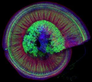
This spiraling organ residing in the mammalian inner ear is responsible for receiving sounds from the environment and transmitting them through neurons (green in the picture, cell bodies in the center) to the brain. The red is phalloidin which binds to the internal skeleton (actin) of the other cells in this organ (hair cells and support cells). The blue is a stain for DAPI, which binds to DNA in the nucleus of each cell.
Developing Motor Neurons and Muscles in E11.5 Mouse Embryo
Courtesy of Jess Bell, Mary Whitman, and the lab of Elizabeth Engle
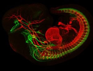
This image depicts a developing mouse embryo with muscles in red, and motor neurons in green.
Multicolor FlpOut clones of Drosophila Ellipsoid Body Ring Neurons
Isabel D’Alessandro, lab of Rachel Wilson
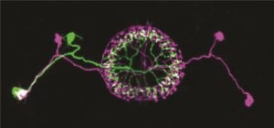
Two ring neurons are labeled on opposite sides of the fruit fly brain. These neurons convey visual information to the brain’s internal ‘compass’ and are important for establishing and maintaining the fly’s sense of direction.
Cerebellar checkers
Ellen DeGennaro, lab of Christopher A. Walsh
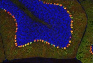
This image shows a thin slice from an adult mouse cerebellum, highlighting blue nuclei, and Purkinje cells in red and green. Our lab is studying a gene that is expressed by some Purkinje cells but has different functions in different parts of the brain.
Olfactory bulb glomeruli
Joseph Zak, lab of Venkatesh Murthy
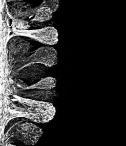
Axon terminals of olfactory sensory neurons terminating in the olfactory bulb at structures called glomeruli. All sensory neurons of a common type project to the same glomerulus and enter as a dense bundle of fibers. Axons are labeled with green fluorescent protein.
News Types: Awards & Honors
